In particular, the “pregnant” part of mule fetus placentas—the uterine horn of the mare that hosts the fetus—tends to have a lower capacity for nutrient, oxygen, and hormone exchange with the dam than that of horse fetus placentas.
However, placentas surrounding fetal mules appear to have a greater capacity for exchange through the nonpregnant parts of the placenta—specifically, the empty uterine horn. While further research is needed to understand the differences, this increased exchange capacity in the rest of the placenta could compensating for reduced exchanges in the pregnant horn, said Claudia Barbosa Fernandes, DVM, BSc, PhD, professor in the Animal Reproduction Department of the University of Sao Paulo’s School of Veterinary Medicine and Animal Science, in Brazil.
Species Differences in the Placentas of Donkeys and Horses
The placenta is a temporary organ that develops from a combination of tissues from both the dam and the fetus. It plays a major role in transporting those nutrients, oxygen, carbon dioxide, and hormones between dam and offspring. Although horses and donkeys have different numbers of chromosomes, hybrid pregnancies are nonetheless possible, in part because donkey and horse placentas share some basic similarities, explained Fernandes.
Despite these similarities, the placentas have important differences, as well. For example, donkey placentas—fetal donkeys gestating inside jennets (female donkeys)—appear to be less efficient than horse placentas, leading to longer gestations and lower birthweights compared to horses.
Surprisingly, however, horse mares carrying mule fetuses don’t have longer gestations, said Fernandes. In fact, they tend to give birth an average of six days earlier than when they’re pregnant with horse foals.
First-Ever Fine-Scale Comparisons of Mule and Horse Placentas
Curious about what’s happening to the placentas in horse and mule pregnancies, Fernandes and her fellow researchers performed the first recorded study comparing the placentas on a microscopic scale.
They collected fetal membranes from nine privately owned Mangalarga Paulista mares, all approximately 10 years old, that had recently given birth to healthy foals. Five of the newborns were horses, and the other four were mules.
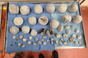
The researchers then spread out each of the mares’ allantochorion membranes—the F-shaped placental structure that in early pregnancy develops and fuses into the endometrium of the uterine body and the two uterine horns, the three compartments of the mare’s uterus. Single fetuses develop in one of the horns, and the other remains empty, but the placenta fills both horn spaces as well as the empty uterine body.
The team photographed and measured each allantochorion membrane and took samples from all the compartments for microscopic analysis and calculations of volume, surface area, and density, among other parameters. The microscopic studies also allowed the scientists to determine the characteristics of each membrane’s microcompartments known as the microcotyledons, the allantoic vessels, the allantoic mesoderm, and the chorion.
Fernandes’ student Julia Boldrini Tinel, DVM, found that each membrane’s weight was about the same—approximately 4.1 kilograms (9 pounds)—for all mares, regardless of whether they had carried a horse or mule foal. That’s significantly heavier than donkey foal membranes, which usually weigh about 2.3 kilograms (5 pounds), she added. Even so, scientists have noticed that membrane weight is usually related to the foal’s body weight; in a healthy pregnancy the placenta is about 11% of the weight of the foal at birth.
The researchers also found that the total volume and the volume of each placental region was about the same for all the mares, with no significant differences between horse and mule foals, she said.
However, the scientists detected distinct differences on a microscopic level between the mule and horse placentas, said Tinel and Fernandes. In particular they noticed variations in the microvilli—tiny fingerlike projections that help connect the uterus (endometrium) and placenta (chorion)—and the microcotyledon—a group of very well-organized microvilli.
For example, in mule pregnancies, the base width of microcotyledons in the empty horn was significantly larger than those of the empty horn in horse pregnancies, they said.
While the findings might seem complex, they narrow down to a more basic general demonstration of what Fernandes refers to as a “compensatory mechanism,” she said. Specifically, the different compartments of the placenta seem to convert and balance out their capacity to manage the pregnancy in different ways, depending on whether the foal is a mule or a horse.
“These differences possibly influence the exchange capacity of each placental microregion and suggest a difference between mule versus horse term allantochorion membrane (meaning the fetus and placenta are mature at birth and the gestational period is complete),” she said.
The study, “ Comparative stereological evaluation of the term allantochorion membrane in the mare pregnant with mule foals and equine foals,” appeared in March 2023 in the Journal of the Animal Reproduction Science.