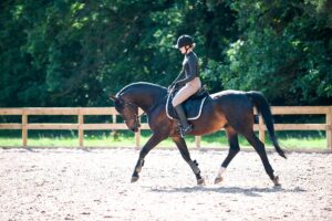
The Systematic Equine Neurologic Exam

Despite best efforts, some cases of equine neurologic disease remain unresolved, without achieving a final diagnosis. The best way to find the answer to your horse’s neurologic issues lies in your veterinarian’s keen eye and methodical approach. A careful history and clinical examination combined with appropriate diagnostic testing help him or her whittle through the long list of potential diagnoses and, ideally, identify the culprit.
Why exactly are neurologic conditions in adult horses so perplexing?
“Horses may be more challenging to diagnose because we are comparatively limited in our diagnostic capabilities, primarily due to size and, in some cases, risk,” says Sarah F. Colmer, VMD, Dipl. ACVIM, a fellow in large animal neurology at the University of Pennsylvania’s New Bolton Center, in Kennett Square.
Kathryn P. Sullivan, VMD, CVA, CVSMT, an assistant professor in New Bolton Center’s Section of Field Service, agrees, adding, “Neurologic diseases in horses are of particular concern due to the size of the animal and the interactions with humans—both in the saddle and in hand.”
In contrast, small animals are much easier and safer to anesthetize and image with computed tomography (CT) or magnetic resonance imaging (MRI).
“Even collecting a cerebrospinal fluid (CSF) sample can be more difficult in the large animal patient from a safety perspective, depending on their degree of ataxia (incoordination),” says Colmer. “Additionally, some relatively common neurologic conditions, like neurodegenerative conditions for example, can only be diagnosed postmortem and, therefore, also present a diagnostic challenge.”
In this article Colmer and Sullivan help us walk rather than stumble through a comprehensive neurologic examination in a methodical, thoughtful, step-by-step manner. Although not specifically addressed in this article, the horse’s history can play an integral role in diagnosing neurologic conditions.
Step 1: Identifying a Neurologic Horse
Not all patients present with classic neurologic signs such as the “dog sitting” of a horse with equine herpesvirus encephalomyelopathy; some cases can have very mild behavior changes and/or ataxia. With mild ataxia, distinguishing between a neurologic and lame horse can be daunting.
“They can be very difficult to distinguish in some cases,” says Colmer, though she notes neurologic disease is often best assessed at the walk, while lameness is best assessed at the trot.
“I typically will briefly trot the horse during a neurologic evaluation and/or put them on a longe line to try to detect if they are lame and, if so, if they are predominantly lame or predominantly neurologic/ataxic versus a combination,” she adds. “Importantly, a significant lameness may hamper your ability to appropriately interpret a neurologic gait evaluation because pain can cause abnormal foot placement.”
Colmer adds that lameness is typically “regularly irregular” whereas ataxia is most often “irregularly irregular.”
“For example, if an abnormal foot placement pattern is consistent and always the same leg in the same fashion and is reliably repeatable, then that might lend itself to more of a musculoskeletal/pain/lameness component,” she says. “If the abnormality is intermittent and/or characterized by a more random foot placement, it may be more ataxic in nature. Importantly, however, horses are not restricted to only one problem.”
“When the clinical signs are mild, which occurs frequently, a work-up typically involves pivoting between multiple diagnostics and imaging modalities,” Sullivan explains.
Step 2: The Physical Exam
Although each veterinarian develops their own pattern for performing a physical and neurologic examination, many elect to start at the nose and end at the tail. Regardless of how they conduct the exam, a main goal is to localize the lesion (i.e., identify the diseased neuroanatomic location) or lesions in one of the following regions of the nervous system:
The cerebrum, or portion of the brain responsible for cognition, mentation, planning action and movement, and integrating sensory information.
The brainstem, which controls many basic life functions (homeostatic mechanisms) and roles such as consciousness, cranial nerve function, and proprioception.
The cerebellum, which plays a role in movement and motor control and coordination.
The spinal cord, consisting of ascending and descending pathways for the transmission of sensory and motor information.
The peripheral nerves originating from the spinal cord that extend to specific regions of the horse’s body, such as the fore- and hind limbs, or from the brain (i.e., the cranial nerves that innervate the head and related structures, carrying both sensory and motor function).
A neuromuscular junction, which is an interface between a nerve and a muscle that initiates and controls muscle contraction and, therefore, movement.

The Horse at Rest
Evaluating the horse’s behavior and mentation or deficits in any of the 12 paired cranial nerves might suggest to the veterinarian there is a lesion within the brain. In some cases mentation changes such as dullness or stupor can increase suspicion for viral encephalitides (e.g., West Nile virus, Eastern, Western, and Venezuelan encephalitis viruses).
Ways to assess the cranial nerves include tongue tone (cranial nerve XII), palpebral or “blink” reflex (cranial nerves V and VII), and eye movement and position (cranial nerves III, IV, VI, VIII), among many others. Some commonly observed cranial nerve deficits in horses include facial nerve paresis (weakness) or paralysis characterized by a droopy lip and/or eyelid on one side of the face, while others include head tilt and dysphagia (i.e., difficulty swallowing).
“Importantly, a cranial nerve deficit does not always mean brain disease,” says Colmer. “Pathology at any point along the tract of the nerve can result in abnormalities. In some cases trauma or a mass may result in compression or other insult to the nerve, resulting in the associated deficits, while the nuclei of the nerves within the brain remain normal. Head tilts can result from otitis (ear infection) or even temporohyoid osteoarthropathy (THO, a bony proliferation of the hyoid apparatus and associated structures).”
“Botulism can also cause difficulty swallowing,” adds Sullivan, providing an example of pathology at the level of the neuromuscular junction, while the brain itself remains unaffected. Intracranial causes of cranial nerve deficits can include equine protozoal myeloencephalitis (EPM), traumatic brain injury, abscesses, and neoplasia (abnormal growths/tumors).
Moving to the horse’s neck and trunk, the main reflexes to evaluate are the local cervicofacial and cutaneous trunci reflexes. The veterinarian performs these assessments by poking the side of the horse’s neck with a pair of closed hemostats (or the tip of a pen).
When the veterinarian taps the neck for the cervicofacial reflex, the horse should exhibit an ear twitch or lip grimace on the side they perform the test. Along the dorsum (back), the horse should exhibit a twitch of the musculature for the cutaneous trunci or panniculus reflexes.
“Testing these reflexes helps us determine at what level of the spinal cord the injury is located,” says Sullivan. “A lack of response may indicate a lesion in the spinal cord that receives sensory input and/or relays motoric output which causes the visible twitch. Compression caused by a tumor or CVSM (cervical vertebral stenotic myelopathy, aka Wobbler syndrome) can impact these reflexes.”
The veterinarian then assesses the perineal reflex (pokes the region with hemostats and observes anal sphincter contraction and/or tail movement) to rule in/out a lesion of either the cauda equina, which is the collection of nerve roots located at the end of the spinal cord near the tail, or the spinal cord sacral segments. He or she might also check anal tone by pinching around the anus.
It’s also important to evaluate muscle atrophy (wasting). “The most common places to appreciate muscle atrophy are the epaxial musculature running alongside the vertebral column and the gluteal musculature,” Colmer says.
A multitude of conditions beyond neurologic diseases such as EPM can cause this atrophy, which people tend to think of most classically. “Nutritional deficiencies and/or poor conditioning can also result in muscle atrophy,” she explains. “The muscle atrophy that occurs with EPM is often asymmetric, whereas atrophy occurring secondary to a nutritional or conditional deficiency is most often symmetric. Horses with deficiency in vitamin E, which is important for neuromuscular health, can exhibit symmetric atrophy.” Some horses can have neurogenic muscle atrophy of one or more limbs due to more focal neuromuscular disease.
“Neurogenic muscle atrophy is typically more dramatic than other types of atrophy, such as disuse atrophy secondary to lameness,” Sullivan adds.
By this point, we’ve assessed mentation, cranial nerves, a few local reflexes, and body condition. What do we know?
- If mentation is abnormal, then a brain issue is likely present.
- If the cranial nerves are abnormal, then a lesion might exist in the brain or along the cranial nerve tract. “But if multiple cranial nerves are abnormal, or if cranial nerve abnormalities are also accompanied by a mentation change, an intracranial (brain) lesion is more likely,” adds Colmer.
- If muscle atrophy is present and asymmetric, veterinarians consider EPM. If it is symmetric, they consider vitamin E or other sources of nutritional deficiency or a lack of work.
The 4 Spinal Cord Segments
The spinal cord is divided into four functional sections. Lesions in each section can affect horses in various ways:
- Cervical (C1-C5) Typically, all four limbs are affected with upper motor neuron signs (longer stride length, increased reflexes).
- Cervical intumescence (C6-T2) Typically, all four limbs are affected, but the thoracic limbs (forelimbs) might be more so. Horses might have lower motor neuron signs (weakness, shorter stride) in the thoracic limbs and upper motor neuron signs in pelvic limbs. The thoracic limb reflexes might be decreased and pelvic limb reflexes increased.
- Thoracolumbar (T3-L3) A lesion here only affects the pelvic limbs, with upper motor neuron signs and increased reflexes.
- Lumbosacral (L4-S1) A lesion in this region affects the pelvic limbs, with lower motor neuron signs and decreased reflexes. Signs include hind-end buckling, decreased anal tone and perineal reflex, and urinary and fecal incontinence.
The Horse in Hand
After completing the exam on the standing horse, watching him move will help the veterinarian further home in on the lesion’s location. This step is particularly important if he or she suspects spinal cord disease and aims to identify which region of the spinal cord is involved.
“Some parts of the brain can (also) be important for gait initiation, maintenance, and coordination, so a gait abnormality is not always indicative of a spinal cord lesion,” warns Colmer.
Veterinarians need to evaluate the gait and look for both paresis and ataxia in each of the four limbs.
“Again, with subtle signs, differentiating between paresis and ataxia can be difficult,” says Sullivan.
When general proprioceptive (spinal, controlling the awareness of body positioning) ataxia is present, both Sullivan and Colmer advise using a grading scale such as the Mayhew modified ataxia scale that ranges from 0 (no ataxia) to 5 (recumbent, or down and unable to get up).
“There are actually three types of ataxia: general proprioceptive, cerebellar, and vestibular,” says Colmer. “Spinal ataxia is the most common and the only one of the three in which we can employ the modified Mayhew ataxia scale.”
Some tests used to determine if a horse is ataxic might include walking in a straight line, tail-pulling while walking, turning in wide circles, tight circles, and serpentines, traversing an incline or decline, walking with head elevation, and reversing. Practitioners watch the limbs for evidence of toe-dragging or overreaching or an otherwise exaggerated gait (hypermetria), as well as a variety of other abnormalities, including delayed protraction (slowness to lift the feet), circumduction (excessive outward swing of a limb), truncal sway, and stumbling and falling.
“We often also watch horses walking up and down hills with their head either neutral or elevated to remove the horizon to see if they place their feet appropriately,” says Sullivan. “Abnormal response would be searching for the ground with their forefeet coming downhill—an almost floating appearance.”
“If a horse is exhibiting general proprioceptive ataxia, then the veterinarian should next determine which region of the spinal cord is affected,” says Colmer.
By this point in the exam, it should now be possible for the practitioner to identify the lesion’s neuroanatomic location. In some cases lesions can be multifocal, making neurolocalization even more complicated. “It can be helpful to write out all of the specific problems and/or abnormalities, neurolocalizing each to the best of your ability,” Colmer says.
Step 3: Additional Testing
Once the veterinarian narrows the list of potential causes for a horse’s condition by neurolocalization, it’s time to pull out the big guns, depending on their top differentials: CSF and bloof analysis, radiography (X rays), myelography, CT, and MRI. CSF can be difficult to acquire on a farm without appropriate equipment, restraint, and technical help, and while the listed imaging modalities can all be useful, their clinical utility might be limited by the sheer size of the horse, local availability of the equipment, and, with some modalities, need for general anesthesia in what might be an unstable patient.
CSF and Blood Analysis
CSF can provide a wealth of information involving cytology, total protein, and disease-specific assays, particularly if the veterinarian’s differentials include infectious conditions. If EPM or Lyme are on that list, paired serum and CSF analyses will provide the most confidence in ruling these diseases in or out, says Colmer. Comprehensive bloodwork might also yield information about potential for infection, electrolyte derangements that can cause neurologic signs, serum vitamin E levels, and liver and kidney health.
X Rays and Myelography
The primary indication for X rays in adult neurologic horses is for cervical vertebral column lesions or head trauma. One of the most common neurologic conditions affecting this region is CVSM, involving narrowing of the cervical vertebral canal and spinal cord compression caused by malformation or malalignment of the cervical vertebrae.
“Regular X rays help either increase or decrease your suspicion of CVSM … but myelography is considered the most sensitive way to diagnose CVSM in the living horse,” says Colmer.
Practitioners perform a myelogram by injecting a contrast dye into the CSF that bathes the spinal cord before imaging the region of interest. Radiographic myelography uses X rays to depict the contrast, but CT myelography is also available at some clinics.
“At the time of this publication, radiographic myelography is generally done under general anesthesia,” says Colmer. “An important consideration for pursuing myelography is the stability of the patient (i.e., if the horse is too ataxic to safely recover, for example).”
Computed Tomography
Next, CT scanning can yield additional valuable information regarding the bony structures associated with the nervous system. It often provides more detailed information in cases such as head trauma. It can also give 360-degree information about the structures of the cervical vertebral column, unlike standard radiography, which only gives us a two-dimensional image, says Colmer. Veterinarians can also use it to evaluate the spinal cord for signs of narrowing. Depending on the unit, CT scans can be performed with the horse under general anesthesia or standing and sedated.
“In some hospitals, CT is their primary method of diagnosing CVSM, but in others it is used in conjunction with radiographic myelogram,” she says.
Magnetic Resonance Imaging
For intracranial abnormalities (e.g., abscess, neoplasia), the test of choice is MRI; however, the patient must be stable enough to transport to an MRI facility (which are limited in number) and healthy enough for general anesthesia. A successful MRI exam can divulge a wealth of information other tests cannot reveal. Examples include intracranial abscessation or hemorrhage, brain edema, neoplasia, or other congenital or traumatic causes.
Take-Home Message
When faced with a neurologic horse, your vet typically conducts a step-by-step exam using a neurologic evaluation followed by advanced diagnostics based on the lesion’s location. Beware that even a thorough exam can fall short, leaving horses undiagnosed in some cases, especially prior to a postmortem.
“Although equine neurology can be very humbling, it is valuable to approach each case methodically, using neurolocalization to guide your differential list, diagnostics, and interventions,” says Colmer. “Consider referring the horse anytime she is a danger to herself or others, having seizures, or the desired diagnostics are not available on the farm.”
| DEGENERATIVE | ANOMALOUS | METABOLIC | NEOPLASTIC | INFECTIOUS/ INFLAMMATORY | TRAUMATIC/ TOXIC |
|---|---|---|---|---|---|
| Equine degenerative myeloencephalopathy/equine neuroaxonal dystrophy | Idiopathic epilepsy | Hyperammonemia (liver, GI, or inborn error of metabolism-associated) | Hemangiosarcoma | Equine protzoal myloencephalitis (EPM) | Toxins such as creeping indigo, arsenic, lead, and many others |
| Cervical vertebral stenotic myelopathy (CVSM, degenerative-joint-disease-associated) | Cholesterol granuloma | Electrolyte derangements (hypocalcemia, -glycemia, -magnesemia, -natremia, hypernatremia) | Lymphoma | Equine herpesvirus-1 myeloencephalopathy (EHM) | Bleeding/hematoma |
| Pituitary pars intermedia dysfunction | Vascular malformation | Osteosarcoma | Rabies | Fracture | |
| Equine motor neuron disease | Peripheral nerve sheath tumor | Bacterial miningitis (Lyme, other) | Ischemia/hypoxia | ||
| Shivers | Eastern, Western, Venezuelan encephalitis viruses | Botulism | |||
| West Nile virus | Tetanus | ||||
| Polyneuritis equi | Edema |

Related Articles
Stay on top of the most recent Horse Health news with


