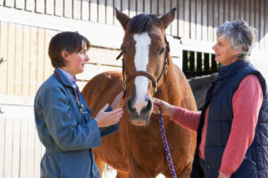
Osteoarthritis can cause neck pain and stiffness in performance horses, leading to poor performance and even a shortened career. Therefore, veterinarians might inject the cervical articular process joints (CAPJ)—where adjacent vertebrae articulate or join one another just dorsal to (above) the spinal cord—with corticosteroids or other therapeutics to alleviate this discomfort. While there are several accepted CAPJ injection methods, Elizabeth Davidson, DVM, Dipl. ACVS, Dipl. ACVSMR, associate professor of sports medicine at the University of Pennsylvania’s New Bolton Center, in Kennett Square, described her preferred technique at the 2023 American Association of Equine Practitioners Convention, held Nov. 29-Dec. 3 in San Diego, California.
Before injecting a horse, Davidson encouraged veterinarians to be certain they are performing CAPJ injections in the right horse, for the right reason, and at the right time. Ideal candidates are horses with authentic, confirmed underlying neck conditions, which are often diagnosed with a thorough exam and imaging. “As with other joint injections, adherence to medication regulations is recommended in the performance horse,” Davidson said.
“CAPJ injections require both knowledge and skill to avoid adverse events,” she added, recommending that veterinarians use an ultrasound-guided technique to optimize success and ensure they are depositing the medication in the right location.
Maximizing Success of CAPJ Injections in Horses
Davidson suggested veterinarians complete the following to increase your chances of success with these injections:
- Refresh your anatomy knowledge before starting and use your hand on the horse’s neck as a rough estimate of one cervical vertebra width. Draw on the horse’s neck with a marker such as white-out correction fluid so you have an idea of where each vertebra is located (topographical anatomy). “Remember, the space between the C5-C6 CAPJ and the C6-C7 CAPJ is smaller than the topographical space between the cranial (towards the head) CAPJs,” Davidson noted.
- Use copious amounts of rubbing alcohol to get good images on your ultrasound. “To identify a CAPJ, you’re looking for a ‘McDonalds sign’—a crescent-shaped area with an echogenic gap (echogenicity shows the density of structures and abruptness of interfaces revealed by ultrasound waves) between the articular processes,” she said. “This ‘M’ arch is a critical ultrasound feature to identify your landmarks.”
- Once you’ve identified the joint, sterilely prepare the skin and sedate the horse. Remember that once sedated, however, the horse will lower his neck, which will change the position of the vertebrae. “Therefore, be sure to prepare a wide area because you may not end up injecting where you originally thought,” said Davidson. For optimal neutral neck positioning, use light sedation only and do not put the horse in cross-ties.
- Use the right needles for your approach and position them correctly. For the cranial approach, veterinarians should use a 9-cm-by-20-gauge spinal needle. Insert the needle 1 to 2 cm cranial to the edge of the transducer, giving yourself some room to reposition needle if needed. Enter the skin at a 30-degree angle in a caudal (toward the tail) direction. You can confirm you are in the right location if you are able to aspirate yellow synovial fluid, she said. “There are other ultrasound clues to confirm you’re in the right location,” she added. “Look for the hyperechoic (appears higher density) shadow of the needle entering the joint. Plus, echogenic specks can sometimes be seen entering the CAPJ during medication instillation, and there should be a lack of fluid in the surrounding soft tissues after injecting.”
Risks and Complications of CAPJ Injection in Horss
At times, the injection process might not be as smooth as described. For example, some pathology in the CAPJ might not allow the needle to enter the joint, such as osteophyte (bone spurs) obstruction. In some cases when repositioning the needle, you might see the tracks from the previous needle passage on ultrasound, which can obscure the image during the current procedure.
Complications following CAPJ injection can include patient stiffness in the injected area, acute synovitis (joint flare) or joint infection. If the vertebral artery running along the ventral (bottom) aspect of the joint is punctured, a hematoma and ataxia (impaired coordination) can occur, or the vertebral canal itself can be punctured, also potentially causing ataxia.
“Two key tips are (1) to know the anatomy and (2) keep the CAPJ in the center of the ultrasound screen, staying dorsal to the spinal canal,” Davidson said.