Positron emission tomography can reveal active processes other diagnostic modalities might miss
On a warm, breezy evening at a research center just outside Sacramento, California, in July 2015, a series of nuclear flashes made equine veterinary history.
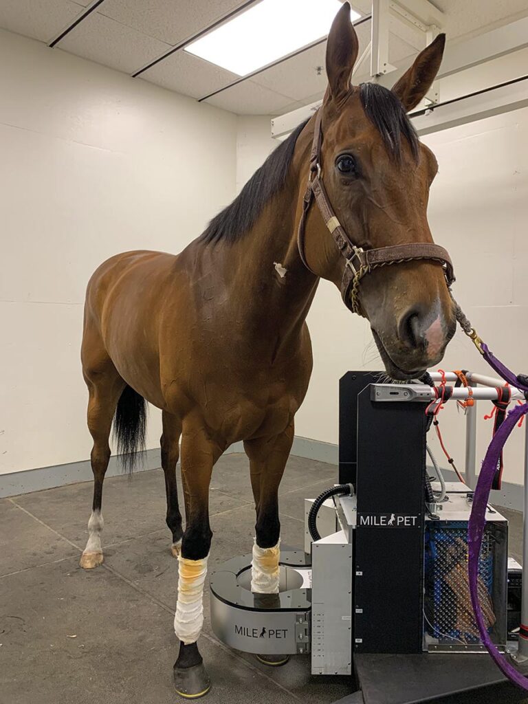
For the previous three months Mathieu Spriet, DVM, MS, Dipl. ACVR, DECVDI, professor of diagnostic imaging at the University of California, Davis, had been experimenting with human brain scanners based on positron emission tomography (PET). At the time doctors mostly used this revolutionary 3D molecular imaging technique to stage cancer in people. Spriet, however, was looking for radioactive glucose uptake in an entirely different target: horses’ limbs.
With only so-so results, though, he considered putting his project on the back burner. That’s when he stumbled across a new paper by researchers trying out another radioactive PET tracer, based on fluoride ions, to diagnose bone pain in people’s feet.
“And I was like, ‘Whoa,’ ” Spriet recalls. “ ‘3D bone scans!’ ”
Shifting gears to the new 18F tracer, Spriet’s eureka moment didn’t disappoint. He scanned the feet of an endlessly and mysteriously lame senior Quarter Horse mare, Fancy Piece of Candy, from the university’s research herd. In a striking moment, a burst of glowing orange radioactivity lit up the ridge at the back of the navicular bone, revealing an active disease process that had not been visible on X ray, computed tomography (CT), magnetic resonance imaging (MRI), and even scintigraphy (bone scan).
“That’s where everything made sense,” he says. “A whole new field of opportunities just opened up for equine imaging.”
Spriet’s radioactive light-bulb moment sparked a rapidly expanding equine PET program at the university—what he affectionately calls “PETting horses.” The approach has since spread across the U.S. and into Europe, giving veterinarians a vivid, real-time view of pain that helps target treatment and detect molecular changes before they can cause catastrophic damage.
Metabolic Maps: Seeing What Other Scans Can’t
In many ways PET represents the new generation of scintigraphy, Spriet says. While these newer tools don’t replace their predecessors—both still play important roles in diagnostics—they offer the key advantage of swapping flat, two-dimensional images for high-resolution, cross-sectional 3D views, allowing clinicians to examine scanned areas slice by slice, in fine detail
This story requires a subscription to The Horse magazine.
Current magazine subscribers can click here to and continue reading.
Subscribe now and gain unlimited access to premium content.
Subscribe NowWe at The Horse work to provide you with the latest and most reliable news and information on equine health, care, management, and welfare through our magazine and TheHorse.com. Our explanatory journalism provides an understandable resource on important and sometimes complex health issues. Your subscription will help The Horse continue to offer this vital resource to horse owners of all breeds, disciplines, and experience levels.