Veterinarians Use MRI to Diagnose Navicular Injury
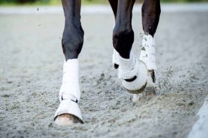
Clinicians at Washington State University recently published a case report about a mare that was referred to the university’s veterinary hospital with chronic left front lameness. The mare’s X rays showed her lame leg was clean, so veterinarians turned to MRI (magnetic resonance imaging), which told a different story–blunt force trauma.
The mare’s owners said she had become acutely lame on her left forelimb while turned out in an arena one day. Radiographs taken by the referring veterinarian didn’t reveal any fractures or fragments, and the mare was placed on stall rest for four months. She was referred to the hospital after failing to improve.
At the university, clinicians performed an MRI scan, which revealed bone density changes in the navicular bone that the radiographs couldn’t identify.
Researchers said, "The most likely explanation is that the navicular bone in the affected limb was injured by blunt trauma, resulting in bone proliferation
Create a free account with TheHorse.com to view this content.
TheHorse.com is home to thousands of free articles about horse health care. In order to access some of our exclusive free content, you must be signed into TheHorse.com.
Start your free account today!
Already have an account?
and continue reading.
Related Articles
Stay on top of the most recent Horse Health news with


