What are the Steps Involved in a Standing MRI?
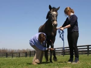
Dr. Rachel Buchholz explains the steps involved in having a standing MRI done on a horse.
Share
ADVERTISEMENT
Dr. Rachel Buchholz explains the steps involved in having a standing MRI done on a horse.
Share
Written by:
Rachel Buchholz, DVM
Rachel Buchholz, DVM, is an associate veterinarian at Northwest Equine Performance in Mulino, Oregon. She currently manages the standing MRI unit at the practice, as well as seeing horses for various lameness and sports medicine related issues. Buchholz graduated from Michigan State University, where she worked in the renowned Mary Ann McPhail Equine Performance Center studying equine spinal anatomy, pathologies, and therapies. Her professional interests include equine physiotherapy, advanced diagnostic imaging, and western performance horse issues.
Related Articles
Stay on top of the most recent Horse Health news with


