diagnosing foot lameness in horses
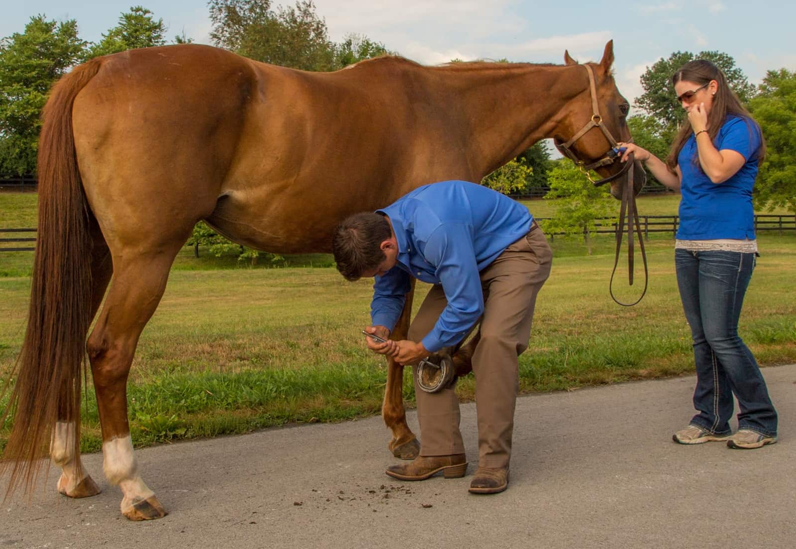
A systematic approach to examining the equine foot can help veterinarians identify causes of lameness more quickly and accurately. To help other practitioners understand the process, Tracy Turner, DVM, MS, Dipl. ACVS, ACVSMR, outlined the steps he’s developed to identify hoof-related pain and imbalances at the 2018 American Association of Equine Practitioners Convention, held Dec. 1-5 in San Francisco, California.

Turner operates Turner Equine Sports Medicine and Surgery, in Stillwater, Minnesota, and has served as an Olympic, World Equestrian, and Pan American games veterinarian over his career.

“During your exam, ask every question three times,” he said. “You will get six different answers, so it’s important to find out before you begin what the goal of your exam is.”

Also, the veterinarian should collect a complete history from the horse’s owner. With an established goal (i.e., why is the vet examining the hoof, and what is he or she trying to find?), Turner described for practitioners his approach to examining cases of equine foot lameness.

Physical Examination

Start at the pastern, assessing the digital pulses, located at the back of the fetlock or pastern region of all four feet. They should be palpable but not bounding like the feeling when you slam your thumb in a car door. The coronet should feel spongy and have a smooth hairline. Appreciate the thickness and density of the collateral cartilages—structures projecting from the coffin bone at the back of the inside and outside of the foot that can become bony (a condition called sidebone). Inspect the hoof wall for cracks, fissures, bulges, uneven growth, heat, or breakage.

“There is always a reason for any observed abnormalities,” said Turner.

For example, the sole should be concave, giving ample clearance between it and the ground; a convex frog might indicate weak and underrun heels. If the frog is receded, it could be because of upright/narrow feet.

Objective Examination

This requires measuring the horse and his foot to assess hoof balance. Collect hoof measurements, including hoof length, heel length, frog length, hoof angle, and frog ratio.

Assess Pain

Watch the horse in motion, Turner said, and see whether his feet land flat or toe-, heel- or quarter- first.

Then, move on to the following tests:

  • A systematic hoof tester examination. A positive response should be repeatable, said Turner—so, if the horse reacts to the hoof testers in a single spot once, he should react every time the practitioner applies pressure. “Using your hoof tests will determine whether there is pain between the prongs or not,” he said. “If so, what’s between those prongs causing the pain? Hoof wall, bone, soft tissues?”
  • Distal (lower) limb flexion. This test puts pressure on structures within the hoof. Hold the limb in question flexed for 30 seconds, then watch the horse trot off. This will reveal 90% of foot pain cases, said Turner, and positive responses often indicate joint or collateral ligament issues. Positive flexion tests might then lead to diagnostic analgesia (joint blocks), diagnostic imaging (e.g., ultrasonography), and/or treatment (e.g., rest).
  • Hoof extension wedge test. Place a wedge under the toe—which places pressure on the navicular bone and bursa—for 60 seconds before watching the horse trot off. Horses that come up unsound could need an ultrasound and might benefit from treatment strategies designed to relieve pressure on the deep digital flexor tendon, which runs from the back of the knee down around the navicular bone.
  • Similarly, the palmar hoof wedge test involves placing a wedge under the palmar (toward the back) two-thirds of the frog/under the heel. Turner said he finds this to be the single best test for pain in the navicular region.
  • Also perform both medial and lateral hoof wall wedge tests, which will put stress on the collateral ligaments.

During any wedge test, pick up the opposite leg, forcing the horse to bear weight on the foot in question.

Take-Home Message

“With every test performed, always ask yourself, ‘What information am I looking for? What imaging modality will provide the information I need?’” said Turner. “Any result from any imaging must always be considered together with clinical findings, and those results will lead to … another test and, ultimately, treatment.”