
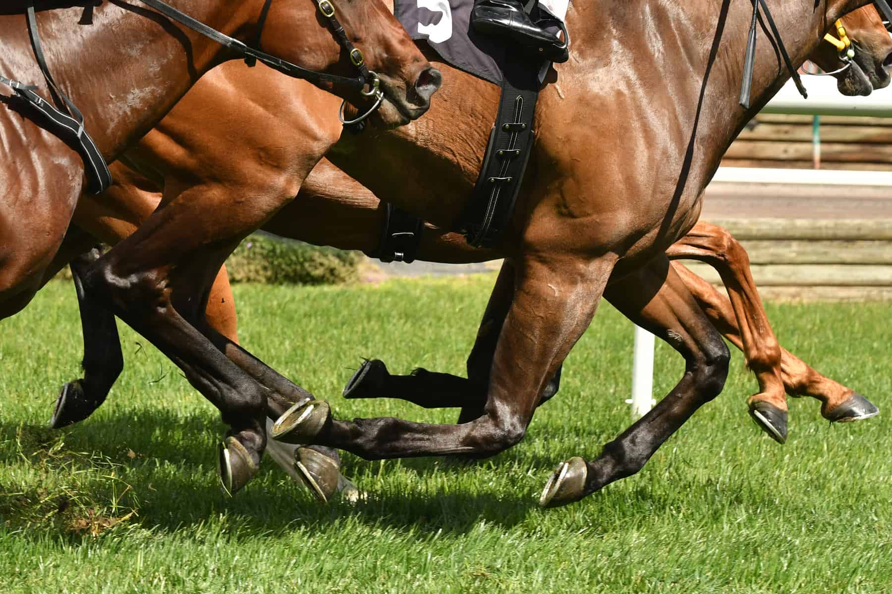
Detecting stress fractures early in is an important part of preventing catastrophic outcomes in equine athletes. But it can be difficult to spot these tiny cracks, especially in a horse’s humerus—the bone between the shoulder and elbow joints—without access to nuclear scintigraphy (bone scan) equipment. But veterinarians recently described a way to do so with a more accessible and affordable approach: ultrasound.
“Humeral stress fractures are well-described in the racing population and can progress to catastrophic fracture if unrecognized,” said Betsy Vaughan, DVM, Dipl. ACVSMR, associate clinical professor of large animal ultrasound at the University of California, Davis, School of Veterinary Medicine.
Nuclear scintigraphy remains the gold standard for diagnosing humeral stress fractures, she said, but it can be expensive and is not available in all locations. Moreover, “radiography can be unreliable to detect stress fractures in the humerus, due to their upper limb location and the time required for sufficient bone remodeling to occur, such that such defects are visible radiographically,” she said.
Ultrasound is readily available to most veterinarians but hadn’t been studied as a tool for diagnosing humeral stress fractures. So, Vaughan and colleagues explored this possibility, and she presented results at the 2018 American Association of Equine Practitioners Convention, held Dec. 1-5 in San Francisco, California.
The researchers reviewed medical records from five Thoroughbred racehorses that were aged 3 to 4 years and suspected of having humeral stress fractures, from June 2013 to February 2018. Vaughan said key findings included:
- All horses with suspected humeral stress fractures had recently returned to the track following recovery from other injuries. They developed severe acute lameness after three to 12 months back on the track, which prompted evaluations for stress fractures;
- In total, treating veterinarians identified seven humeral stress fractures—two horses had bilateral fractures (one in each of their forelimbs), two had fractures on the right, and one had a fracture on the left;
- Ultrasound revealed step defects (a small step or gap in the otherwise smooth bone contour, 3 cases), bone callus or roughening (7 cases), and/or a convex contour of the normally straight bone surface at the back of the humerus (7 cases);
- Both radiography and nuclear scintigraphy revealed signs of stress fracture in seven and three cases, respectively; and
- Repeated re-evaluations with radiography and ultrasonography in two cases showed progressive healing.
As of her presentation, Vaughan said two horses were still being rehabbed, two had returned to their intended uses as riding and racehorses, and one had retired.
In summary, she said, “Ultrasound is a useful and economical screening tool to identify humeral stress fractures and can be used with radiography to monitor healing. It is very important to remember that a normal ultrasound exam does not rule out stress fracture. Radiography and/or nuclear scintigraphy are sometimes required to identify fractures and should be pursued in suspect cases with normal ultrasound findings.”