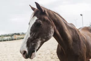
If you look at sensitive hoof tissues of a horse with septic laminitis and one with endocrinopathic laminitis under a microscope (called histology), you can tell the cases apart. Mechanical failure of the laminae, which suspend the coffin bone within the hoof, occurs in both, but it happens differently. In septic cases (with a toxic cause) inflammation and separation of the laminae are evident. In cases linked to endocrine problems, there’s stretching and increased cell proliferation, but no inflammation. These differences are helping scientists understand exactly how and why endocrinopathic laminitis occurs and in which horses.
Nicola Menzies-Gow, MA, VetMB, PhD, Dipl. ECEIM, Cert EM (internal medicine), MRCVS, senior lecturer in equine medicine at the Royal Veterinary College, in Hertfordshire, U.K., has devoted her research career to studying insulin resistance and equine laminitis. She described what scientists currently think happens in endocrinopathic laminitis cases—specifically those associated with EMS—and why during a series of laminitis presentations at the 2016 British Equine Veterinary Association Congress, held last fall in Birmingham, U.K.
The Horse-Human Metabolic Syndrome Conundrum
What researchers recognize as EMS has evolved slightly over the past 10-15 years; they now define it as a cluster of clinical and metabolic abnormalities that are associated with an increased risk of laminitis.
“It’s not these abnormalities equal laminitis, it’s an increased risk,” said Menzies-Gow. “And the (initial EMS) definition has come from human metabolic syndrome, I think that’s where we suffer a little bit when it comes to EMS. We’ve assumed because that if it happens in people like this, then it happens in horses like this, and that’s not always the case.”
In human metabolic syndrome cases, physicians see obesity (particularly visceral—or fat accumulation around the organs in the abdomen), insulin dysregulation, hypertension (high blood pressure), an abnormal plasma lipid profile, a chronic low-grade inflammatory state, and altered hormones that are associated with adipose, or fat, tissue (called adipokines). These clinical signs put patients at an increased risk of suffering from cardiovascular disease and Type 2 diabetes.
Equine metabolic syndrome horses, on the other hand, could be obese, but they don’t have to be. Also, researchers doubt that EMS horses live in a chronic inflammatory state or with hypertension, Menzies-Gow said. Rather, “The consequence for the (EMS) horse is an increased risk of laminitis.”
Overall, key features of EMS are still evolving, particularly because past studies have included horses of different breeds, ages, and sexes and kept in slightly different environments, which can confuse researcher agreement. Menzies-Gow said the current working key features are:
- Insulin dysfunction;
- Increased fasting triglyceride concentrations (a type of fat in the blood); and
- Altered adipokine concentrations.
Essentially, horses matching this profile are primed and ready for a clinical laminitis episode, commonly triggered by grazing pasture. “Our current working thoughts are that with consumption of this carbohydrate (fructans) you get a pronounced insulinemic response, which then tips the pony over that threshold and into overt laminitis,” said Menzies-Gow.
She offered the alternative theory that the carbohydrate instead enters the hindgut (the gastrointestinal tract beyond the small intestine), triggering a carbohydrate overload mechanism and absorption of trigger factors, though she favors the hyperinsulinemic response theory.
Breed Predilection
“Not every pony that has these metabolic abnormalities that goes out to pasture will get laminitis every time,” she said, “so there must be some kind of alternative influence that’s making the individual animal end up being laminitic, whereas the identical one standing next to it grazing exactly the same pasture doesn’t get it.”
That puzzle’s answer is genetics, said Menzies-Gow, because when you consider horse breeds more likely to get EMS, there’s definitely a “type.”
[image imageid="6670" includeTitle="false"][/image]
“There are certain breeds that appear to be overrepresented,” she said. “We all know it’s the U.K.-native pony breeds, it’s the Andalusians, it’s things that they get in the (United) States, the Tennessee Walking Horses … Morgan horses, so there’s some kind of breed influence.”
Horses and ponies have genetic alleles (variants) that influence their insulin production and their glucose and lipid metabolism, some of which could decrease their risk of laminitis, she said, and others that increase it. “And those work together with your environment (think sugar levels in grasses, exercise levels, etc.) to give you your overall risk.
“There are some things we can influence, particularly the environment,” she said, referring primarily to dietary management, “but obviously you can’t change the pony’s underlying genetics.”
The Mystery of the Stretching Lamellae
So, how does EMS give you laminitis? Of the three laminitis research models scientists use (carbohydrate overload, black walnut toxicosis and infusion of high doses of insulin), it’s important to look at the insulin infusion model, because that one causes hyperinsulinaemia without inflammation, much like EMS does.
Initially researchers thought EMS-rooted laminitis was a glucose problem—either glucose deprivation or glucose toxicity—because study horses’ high insulin levels indicated they were unable to get glucose into the cells for storage as glycogen. “But we’ve since discovered that glucose transporters that are in the hoof bed are not insulin dependent,” she said. “So that this is not the underlying cause.”
The current theory is that insulin causes cells in the blood vessel lining (endothelial cells) to produce too many vasoconstrictor mediators and not enough vasodilator mediators. In other words, blood vessels constrict rather than dilate, restricting blood flow. In the vital tissues of the hoof, that would result in laminitis.
“Another effect of too much insulin on the blood vessels is it promotes platelet and leucocyte adhesions (basically, platelets sticking to the white blood cells), both of which are a feature of laminitis,” she said.
But Menzies-Gow said the mechanism that’s getting the most attention right now has more to do with cell signaling than blood vessels. Insulin released in the body at normal levels binds to insulin receptors (InsR). At higher levels, the insulin can also bind and activate the insulinlike growth factor-1 receptor (IGF-1R) and/or a hybrid of the two (InsR/IGF-1R). Researchers believe this could weaken the lamellar suspensory apparatus—that characteristic stretching seen on histology—and trigger the onset of laminitis clinical signs.
“It certainly seems that the IGF-1R receptor is the predominant receptor type in equine lamellar tissue, and it’s responsible for regulating cell growth and cell adhesion and survival,” she said. “So it might be that … hyperinsulinemia is inappropriately stimulating IGF-1R, stimulating the epidermal cell proliferation, and that is the key feature that we see in the histology of these models. But it’s yet to be completely determined.”