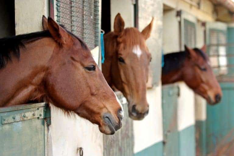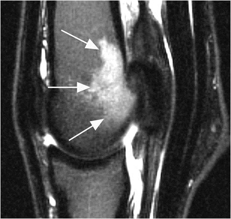
Using MRI to Evaluate Fetlock Pain
Vets use MRI to identify issues and prescribe targeted treatment to give the horse the best chance at returning to work.

Vets use MRI to identify issues and prescribe targeted treatment to give the horse the best chance at returning to work.

Researchers found that meropenem can be useful, but should only be used when other antibiotics have failed. Here’s why.

While not frequently diagnosed, equine muscle injuries can cause pain, lameness, and poor performance in horses.

Horses with exertional myopathies can benefit from dietary modifications as well as consistent targeted exercise.

Thermography is a noninvasive, safe, and cost-effective diagnostic imaging tool used in equine health care.

One researcher describes the promising progress that’s been made in lameness diagnosis and treatment.

Vets can glean crucial information by evaluating horses with performance issues in hand, on the longe, and under saddle.

Researchers found that MRI images of bone thickness could provide critical information about fracture risk.

Jasmin Bagge, DVM, is studying the use of stem cells to facilitate tissue repair following injury in horses.

Of the 442 respondents, 90 (20%) use injections as a regular part of their horses’ joint management programs.
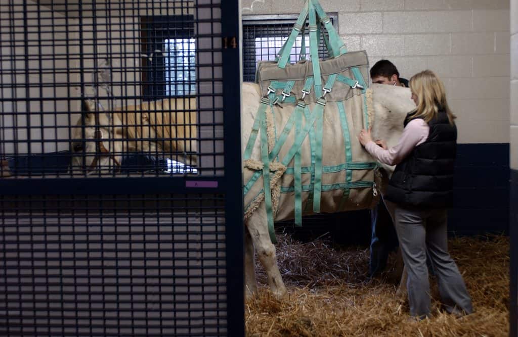
The University of Wisconsin School of Veterinary Medicine used donated funds to purchase the new equipment.

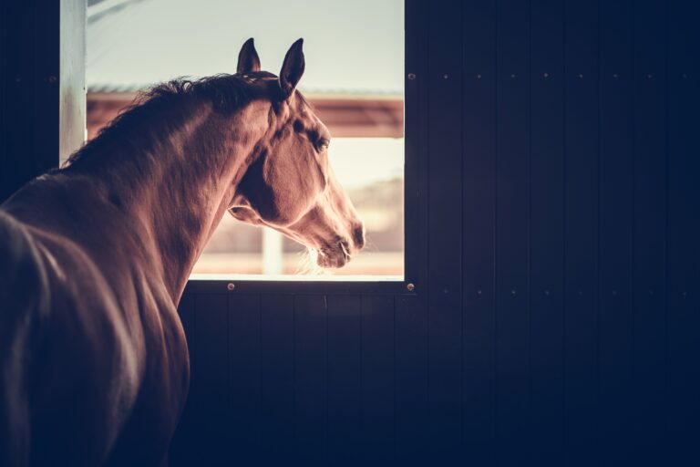
Veterinarians use imaging technologies to evaluate, diagnose, and ultimately treat lameness in the lower leg and foot.

Complications are common with traditional repair, so researchers tested a plate designed for human geriatric patients.


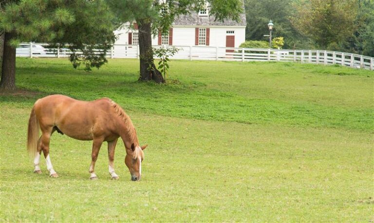
Learn how recreational riders can condition and protect their equine weekend warriors.
They might be less common than limb fractures, but skull, rib, pelvis, and withers fractures are no less important.