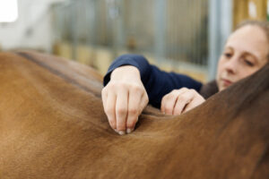
So how can vets use what they’ve elucidated from MRI findings to better identify and diagnose radiographic lesions? Marianna Biggi, DVM, PhD, FHEA, Dipl. ECVDI, MRCVS, head of the equine group at Vet-CT and clinical radiologist at Pool House Equine Clinic, addressed this topic during a presentation at the 2021 British Equine Veterinary Association Congress, held Sept. 4-7 virtually and in Birmingham.
“MRI has helped not only to recognize ‘new’ pathologies within the foot, mainly affecting the soft tissues, which were previously going undiagnosed, but it has also improved our understanding of the clinical significance of some well-recognized radiographic findings and the association between bone and soft tissue pathology within the foot,” she said. “We’ve recognized these lesions for a long time, but we know better what they mean based on what we’ve learned from MRI.”
Shining Light on Known Lesions
Biggi briefly described lesions veterinarians have become better at interpreting radiographically thanks to MRI:
Navicular bone lesions
Since MRI’s introduction, veterinarians have discovered the navicular bone is rarely the only affected structure within the podotrochlear apparatus. Further, “comparing MRI and radiology of the navicular bone has revealed that subtle radiological changes may be indicative of severe pathology on MRI and that a normal radiological study does not exclude severe navicular bone disease,” she said.
For example, she said, MRI has improved veterinarians’ understanding of changes to the distal edge of the navicular bone, such as osseous cyst-like lesions and fragments, that many previously considered incidental.
Capsulitis (joint capsule inflammation) at the insertion of distal interphalangeal (coffin) joint
Biggi said veterinarians often underestimate the significance of this change on radiographs, especially if a horse went acutely (suddenly) lame. “You see these new bone formations that look quite chronic and thickening of the dorsal cortex (of the middle phalanx where the joint capsule attaches),” she explained. “Many of these horses have increased signal intensity on the cortex itself, and they don’t necessarily block to the intra-articular block. But this is a clinically significant finding.”
Ossification (hardening) of the ungular cartilage on either side of the coffin bone
Biggi said back when she was in veterinary school, this was an incidental finding. “Now we know it’s not, especially if we see cartilages that are thickened and have increased opacity (on radiographs),” she said. “These will translate to sclerosis (denser than normal bone) on MRI and potentially bone edema (fluid within the bone).”
Biggi said MRI has also shown veterinarians a world of chondral ligaments and ligaments that connect the cartilage to the other bones within the foot that are difficult if not impossible to diagnose using radiographs alone.
What Have We Learned From MRI?
Biggi summarized the main things MRI has taught veterinarians about foot lesions:
- Subtle radiological changes in specific locations can represent severe pathology on MRI, especially in known locations, such as the palmar compact bone of the navicular bone.
- Subtle radiological changes are more difficult to interpret and identify in poorly positioned radiographs, making well-positioned, clear radiographs a must.
- Multiple injuries usually coexist within the foot. “Even if we do find a lesion we think might be significant, there might be much more going on within the foot, and it’s still worth sending the horse for an MRI to have the exact extent of pathology and give the correct treatment and prognosis,” said Biggi.
- Extensive pathology can be present on MRI despite normal radiographs, and slight changes in opacity on radiographs might show up as significant bony lesions on MRI. “There are radiographs that are completely normal and we’re not missing anything, but this does not preclude the presence of extensive pathology on MRI,” she said.
While Biggi recommends careful interpretation of a well-positioned and complete set of radiographs before pursuing advanced imaging techniques, MRI remains a crucial tool for diagnosing foot lameness in many cases.