How coffin bone fractures happen, and how to prevent them from becoming career-ending injuries
The coffin bone, also called the pedal bone or distal phalanx, is the terminal bone in the horse’s limb, encased within the hoof capsule. It serves as the foundation of the foot, where important structures ranging from the deep digital flexor tendon to the laminae attach. Despite its location behind the protective hoof wall, the coffin bone can fracture, resulting in one of seven types of breaks.
“Picture hitting an icicle with a baseball bat. If you swing like you are trying out for the Phillies, the ice shatters into a million pieces,” says Janik Gasiorowski, VMD, Dipl. ACVS, associate veterinarian and equine surgeon at the Mid-Atlantic Equine Medical Center, in Ringoes, New Jersey. “But if you were to push that same baseball bat slowly into contact with the icicle, it would only break into two clean pieces.”
The same holds true for bones. “All fractures happen quickly, but if you pause and ‘zoom in’ on the moment of impact, you see that the faster the force is applied, the more energy is stored in the bone, and the more violently that bone breaks,” Gasiorowski continues. “This difference accounts for ‘good’ versus ‘bad’ outcomes, with shattered bones carrying a worse prognosis than clean breaks.”
Indeed, shattering is a sign of high energy input at the time of fracture. “When the coffin bone is shattered into many pieces, we understand the energy input clearly because we see the resulting bone fragments,” he explains. “But things are not always so clear-cut.”
A coffin bone fracture can come with myriad invisible insults to surrounding structures. “Sometimes, much of the excess energy is absorbed by the soft tissues—cartilage, joint capsule, ligaments,” says Gasiorowski. “We cannot see squashed cartilage or torn ligaments on radiographs. Over time, these injured tissues scar or die. Damaged hyaline cartilage (the glassy smooth cartilage that lines joint surfaces) in the coffin joint gets replaced with (more rigid) fibrocartilage; the joint capsule and ligaments get thicker and stiffer. These changes cause arthritis.” Arthritis, as we’ll discuss in this article, can cast a dark shadow over an otherwise bright outlook for horse owners rehabbing a fracture.
Causes of Coffin Bone Fractures
If you consider the unique anatomy of the equine athlete, it could seem surprising that coffin bone fractures are a rarity rather than an inevitability. A half-ton animal landing at great speeds and with great force upon a single hoof at a time at the canter and gallop places a tremendous amount of pressure on the coffin bone.
Veterinarians and researchers (Kidd 2011, Morrison 2013) have found the most common causes of a broken coffin bone include:
- Trauma, which accounts for most fractures of the equine digit, such as kicking or colliding with firm objects, or racing injury.
- Improper shoeing, leaving the coffin bone to bear more weight and pressure than nature intended.
- Working on hard surfaces, inflicting repeated concussion on the bone.
- Stone bruises.
- Infectious conditions inside the hoof capsule, which can progress to osteomyelitis (bone infection) and fracture.
- Nutritional deficiencies—notably calcium and vitamins A and D—which weaken the coffin bone and predispose it to fractures.
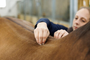
Regardless of the type of coffin bone fracture, the most apparent clinical sign is generally lameness. To make a diagnosis veterinarians perform physical and lameness exams, including the application of hoof testers and possibly nerve blocks to locate the source of pain. Radiography (X ray) is the veterinarian’s go-to diagnostic imaging modality for suspected fractures.
Christina Cable, DVM, Dipl. ACVS, owner of Early Winter Equine, an ambulatory practice in Lansing, New York, sees three to four coffin bone fractures per year, most of which are wing fractures. “The fractures tend to be in the forelimbs, but very occasionally we find them in the hind limbs,” she says. “This is likely reflective of the distribution of the horse’s body weight, with an estimated 55-60% carried by the front end and the remaining 40-45% by the hindquarters. With more weight comes a greater risk of injury.”
The Challenges of Joint Damage
Not all fractures are created equal, mainly because bones and joints are not on a level playing field when it comes to healing. On one hand, bone can be 100% fixed and return to its previous strength. “As a matter of fact, bone heals very well and continues to remodel for years after primary healing, adapting to daily use based on the stresses applied to it,” says Gasiorowski. “Even when a fibrous union (not true bone healing) occurs in nonarticular fractures, the horses are usually sound and comfortable.”
Joints? That’s a different story, which is why articular coffin bone fractures are bad news. “Cartilage has very limited capacity for healing,” says Gasiorowski. “The main concern with articular fractures is the development of arthritis. More specifically, hyaline cartilage is the problem. Hyaline cartilage can heal small defects (from normal wear and tear), but sustaining significant trauma from a fracture can cause permanent damage. The hyaline cartilage of the coffin joint is destroyed and replaced by fibrocartilage. Fibrocartilage, as its name indicates, has a fibrous consistency. It lacks the tensile and expansile properties of hyaline cartilage, making it an inferior substitute. Fibrocartilage also does not form with the exact same shape as the hyaline cartilage originally had, leaving bumps and divots in the gliding surface of the joint.”
Researchers recognize arthritis as the main complication of coffin bone fractures. In a 2021 study Smanick et al. found an 86% return to work rate with surgical repair of the fracture and confirmed that arthritis was the complication with the greatest negative impact on success. What does that mean for the horse’s athletic career? Essentially, attempting to rehabilitate a horse with trauma-induced arthritis could reveal limitations.
“At rest, a joint ‘patched up’ with fibrocartilage may be just enough to keep the horse comfortable, but it breaks down under the more intense loading experienced during athletic use,” says Gasiorowski. “The incongruency of the joint surface and the constant breakdown and reinjury of the fibrocartilage create an environment of persistent inflammation within the joint. This leads to the production of catabolic enzymes (chemical substances that break down the joint) and poor-quality, watery synovial fluid (the lubricating and nourishing fluid of the joint). This combination of changes results in damage to the articular surface and joint capsule even well beyond the location of the initial injury.
“We can treat to some degree the inflammation, but we cannot reverse the damage or remove the cause of these problems—namely, inferior cartilage and joint incongruency,” he continues. “So things continue to spiral downward in the degenerative process we know as arthritis. We have limited ability to help these arthritic joints with various medications and joint injectables (see sidebar on page 64). Unfortunately, the only way to truly slow degeneration is to decrease use: stop racing, jump lower, or even retire.”
Surgical Management
“Thoroughbred and Standardbred racehorses overwhelmingly comprise our caseload for distal phalanx fractures, partially because their sport predisposes them to this type of injury,” says Gasiorowski. He says he’s firmly in favor of repairing articular fractures surgically. “In contrast to earlier recommendations, I’ve found through current studies and my personal experience that with articular fractures of the coffin bone, the majority of horses with surgical repair return to racing, whereas those without surgery do not.”
Looking at the rate of return to racing both in the 2021 paper mentioned and his own surgical caseload, Gasiorowski finds it difficult to support nonsurgical management of articular fractures of the distal phalanx. “Very young horses are an exception,” he notes. “They do well with a shoe and rest alone. Other practitioners have suggested nonsurgical management for horses less than 3 years of age, but I believe that cutoff should be one year, especially when talking about racehorses.”
So, the need for surgery depends on three main factors:
- The nature (type) and extent of the fracture, with Types 2 and 3 being the most common surgical candidates.
- The age of the horse.
- The owner’s goals for the animal.
“Horses that develop coffin bone fractures with articular involvement often require surgery in order to minimize the development of osteoarthritis,” says Cable. “We also use strict stall rest and (therapeutic) shoes with multiple clips to help try to stabilize the fracture and prevent any joint movement.”
Of course, the potential of arthritis developing is a big consideration when deciding whether to operate. “There’s no doubt that internal fixation (surgical repair) can greatly reduce the risk of arthritis developing in an articular coffin joint fracture,” says Gasiorowski. “The distal interphalangeal (coffin) joint is a high-motion joint; prevention of arthritis is critical. These fractures are repaired with one or two screws placed across the fracture in lag fashion where the screw(s) squeeze the two pieces together. The goal is to eliminate motion at the fracture gap and stabilize the bone. This results in better-quality healing: Alignment at the joint surface is maintained, and the bone heals directly instead of with callus. These principles are important in all fracture repair but especially in a joint.”
Bone movement and fracture instability are major instigators of inflammation, degeneration, and osteoarthritis. “Surgical repair takes care of that,” he notes.
Post-surgery, he says, horses go on stall rest for one to two months, graduate to small turnout or light active rehabilitation for two to three months, and can resume training in four to five months, if all goes well. The screws typically remain in the bone.
Final Thoughts
Arthritis is the key factor to consider when determining a horse’s prognosis after distal interphalangeal fracture. “If arthritis develops from a coffin bone fracture, the horse will be a lesser athlete or not an athlete at all,” says Gasiorowski.
Predicting the onset of arthritis and acting quickly, often with surgical intervention—which allows nearly nine of 10 horses with coffin bone fractures to return to work—gives horses the best chance at a pain-free, healthy joint and life.