Veterinarians Evaluate Adverse Reactions to Myelograms

“For the most part, the reactions were mild and self-limiting,” said study author Kathleen Mullen, DVM, MS, Dipl. ACVIM, an instructor in equine and farm animal internal medicine at Cornell University, in Ithaca, New York.
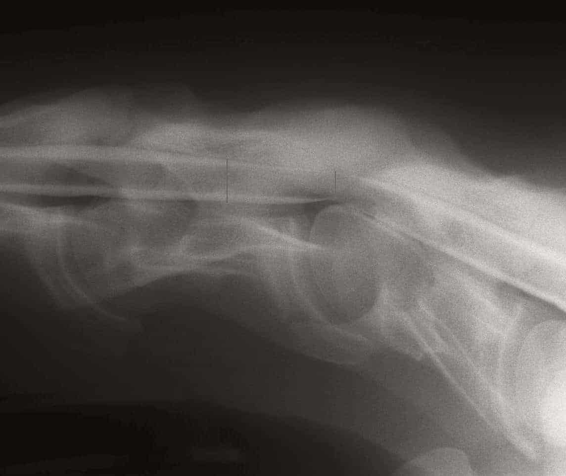
During a cervical (neck) myelography, the veterinarian carefully inserts a spinal needle into the space between the skull and the first vertebrae (called the atlanto-occipital space) and removes a small amount of cerebrospinal fluid (CSF) for analysis. Then, he or she injects contrast material and takes neck radiographs.
In normal horses, the contrast should be evident along the entire length of the spinal cord in the neck, even when the neck is flexed or extended. A horse with a compressive lesion, however, will have less contrast where the spinal cord is compressed. Vertebrae deformities, developmental bone disease, osteoarthritis, trauma, or masses can cause cervical spinal cord compression in horses
Create a free account with TheHorse.com to view this content.
TheHorse.com is home to thousands of free articles about horse health care. In order to access some of our exclusive free content, you must be signed into TheHorse.com.
Start your free account today!
Already have an account?
and continue reading.
Written by:
Katie Navarra
Related Articles
Stay on top of the most recent Horse Health news with


