A look back at the incredible amount of research scientists published about osteoarthritis in the past year.

It’s been a busy year in equine osteoarthritis research. Over the past 12 months scientists have published approximately 100 papers in academic journals on their discoveries about this debilitating inflammatory joint disease in horses.
Representing up to 60% of all lameness cases, osteoarthritis (OA) is a whole-joint disease characterized by inflammation, cartilage breakdown, and bony lesions and/or other changes in subchondral bone (which lies directly beneath the cartilage). While OA affects the entire joint, cartilage destruction, in particular, represents the “central and irreversible step in the process” of the disease, says Raquel Baccarin, DVM, PhD, professor and head of the equine internal medicine service at the veterinary hospital at the University of São Paulo, in Brazil.
Cartilage breakdown occurs when cartilage cells—called chondrocytes—no longer produce and secrete proteins that build the extracellular matrix and keep the cartilage structure strong. Instead, the chondrocytes secrete inflammatory cytokines or short RNA fragments known as microRNAs (miRNAs) that cause cells to degenerate or die.
Diseased cartilage is challenging to treat because cartilage has no blood vessels, meaning the body can’t transport healing agents into its cells, Baccarin says. Plus, chondrocytes rarely replicate and, therefore, replace dead or damaged cells.
Eager for better understanding, detection, prevention, and management of equine osteoarthritis, scientists across the globe have been investing significant time and effort into studying the disease. In this article we’ve delved into the studies published in the past year to provide you with an essential rundown on what’s new.
Osteoarthritis Prevention, Diagnostics, and Monitoring
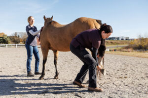
Prevention, early diagnosis, and close monitoring of disease progression are key factors in dealing with an irreversible disease such as osteoarthritis, our sources say, so scientists are actively researching these areas.
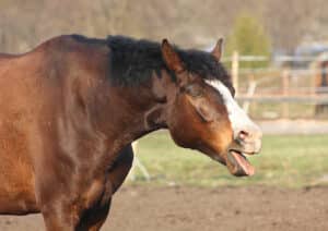
One clue could be subtle clinical signs, says Eva Skiöldebrand, PhD, professor at the Swedish University of Agricultural Sciences, in Uppsala. Horses might first appear stressed or depressed, or they might be reluctant to move forward.
Scientists in Switzerland and Canada discovered when horses had arthritic inflammation in a jaw joint, they became more sensitive to rein tension on that side and needed more rein tension on the other side to keep the head straight. And researchers at the University of Kentucky found reduced muscle mass was a sign of osteoarthritis in senior horses.
Also in Switzerland, scientists investigated the center of pressure paths (COPp) in horses’ feet, which are as unique for each horse as fingerprints are for humans. Using sensors and a wireless pressure measurement system, the team determined the COPps in lame feet take a very different trajectory than those in the sound feet of the same arthritic horses. Comparing normal COPps for each horse’s feet to any subtle changes could signal early effects of osteoarthritis, the researchers say.
As osteoarthritis advances, cartilage becomes thinner, so researchers at the University of Copenhagen, in Denmark, explored ways to monitor cartilage thinning using ultrasound. Comparing different ultrasound techniques to MRI and laboratory measurements on multiple sites of eight Standardbreds’ knees, they determined that a 15 MHz—but not a 9 MHz—ultrasound transducer can reliably measure cartilage thickness.

Cartilage damage might even start in subchondral bone, say New Zealand researchers. It can stiffen and compromise the entire joint’s shock absorbency, leading to physical damage to the overlying cartilage. So it makes sense to investigate subchondral bone integrity with imaging, they note. In their study using polarization-sensitive optical coherence tomography (PS-OCT), they successfully detected microscopic bone changes in five cadaver limbs that would otherwise have been impossible to see without dissection.
Meanwhile, at Michigan State University, in East Lansing, researchers are questioning whether horses—like people—might be developing what’s known as “metabolic osteoarthritis” provoked by chronic low-grade systemic inflammation related to endocrine disorders such as equine metabolic syndrome (EMS). The team has studied this possibility and recently published a paper encouraging further investigation.
As for management strategies aimed at preventing osteoarthritis—which often include various trimming and therapeutic shoeing techniques, compression bandaging, and nutraceuticals, with the goal of minimizing damage to joints—there’s little evidence they work. Researchers at the University of Liverpool, in the U.K., recently reviewed 10 years’ worth of studies and found a lack of robust research to support these methods. The team emphasizes the need for well-designed investigations into these management strategies, says Emily Clarke, a PhD candidate in the lab of Mandy Peffers, PhD, FRCVS.
Biomarkers
The lion’s share of recent research on equine osteoarthritis has focused on biomarkers—and for good cause. Biomarkers can pick up osteoarthritis early enough to manage the disease before the horse feels pain, says Skiöldebrand.
“By the time you see changes on X ray, the horse has had the disease for a long time, perhaps many years,” she says. “But with biomarkers, we can actually monitor the exact phase when young horses’ bone and cartilage start to degrade.”
For the past decade scientists have been seeking these biomarkers in horses’ blood (serum or plasma), saliva, or synovial fluid. Synovial fluid is lubricating liquid produced by the synovial membrane that surrounds each joint. Synovial fluid biomarkers point to disease in specific joints because the veterinarian punctures a joint to aspirate its fluid, whereas saliva, serum, and plasma biomarkers point to osteoarthritis somewhere in the body.
Recently, Italian researchers used proton nuclear magnetic resonance (H-NMR) spectroscopy to discover that fetlock synovial fluid in arthritic horses had more 1,3-dihydroxyacetone but less tyrosine, uridine, creatinine, creatine, glycine, and 13 other biomolecules compared to healthy horses. The team suspects the joint’s metabolome—the complete set of small-molecule chemicals found within the sample—gets out of balance in arthritic horses as it tries to cope with inflammation, they say.
Inspired by studies in humans, scientists at Cairo University, in Giza, Egypt, found that donkeys had increased expression of the genes miR-146b and miR-27b in serum and of the Col10a1 collagen gene in synovial fluid during the early stages of knee arthritis, suggesting in their study that they’ve identified “promising noninvasive biomarkers for the very early diagnosis” of osteoarthritis. U.K. researchers, meanwhile, found another short RNA gene, miR-92a, was downregulated in the synovial fluid of horses with severe osteoarthritis.
At Massey University, in New Zealand, Luca Panizzi, PhD, and his colleagues detected statistically higher concentrations of cell-free DNA (cfDNA) in the synovial fluid—but not the serum or plasma—of arthritic Thoroughbred fillies than in healthy ones. Their fourier-transform infrared (IR) spectroscopy—a rapid scanning technique known for identifying the “biochemical fingerprints” of all molecules in biofluids—couldn’t detect osteoarthritis biomarkers in these horses’ serum samples.
Meanwhile, scientists at the University of Eastern Finland, in Kuopio, found significantly fewer large-sized extracellular vesicles—the “vehicles” that transport certain substances between tissues—in osteoarthritic horses’ synovial fluid. These are the vesicles capable of transporting the natural lubricant, hyaluronic acid, reports Anne-Mari Mustonen and her colleague. They also found that the synovial fluid of osteoarthritic fetlock joints contained far more 18:2n-6, a fatty acid derived mainly from nuts, seeds, and oils but also hay and oats. This suggests the lipid might serve as a biomarker, “but it would also be worth investigating if 18:2n-6 could be a contributing factor to arthritis,” she says.
U.K. researchers discovered seven short RNA genes within the extracellular vesicles of plasma and synovial fluid that could serve as biomarkers for early osteoarthritis, as well as four small RNAs of a different category, known as nucleolar RNA.

Suspecting native biglycan (BGN)—a protein involved in bone formation and mineralization—might be implicated in osteoarthritis, Skiöldebrand and her colleagues screened synovial fluid for a cleavage product of BGN, known as BGN262, associated with inflammation in the bone matrix. In their study of 88 horses, they found significantly increased concentrations of BGN262 in the synovial fluid of joints with more severe subchondral bone sclerosis—a suggested precursor to osteoarthritis. They also noted increased concentrations of BGN262 in horses during their first six months of racetrack training and a tremendous increase in those with chip fractures in the joint, she says.
In another study her team found a synovial fluid biomarker, a cleavage product of cartilage oligomeric matrix protein called COMP156, which was detectable in cases of acute lameness associated with osteoarthritis, she says.
Together, the biomarkers can reflect the joint’s ongoing health status, she explains.
Pro-inflammatory immune cells such as T helper cells could serve as biomarkers to determine disease severity. A team at Cornell University aspirated synovial fluid from horses with and without post-traumatic arthritis and found—for the first time in horses or humans—that in more severe cases T helper-17 (Th17) cells outweighed the natural anti-inflammatory Treg cells, effectively upsetting a healthy Treg-to-Th17 balance. The findings might help guide therapeutic approaches after joint injuries, the researchers report.
Biomarkers could also help pinpoint which arthritic horses are in pain, Skiöldebrand adds. In an initial study of 69 horses, her team’s findings suggested high serum concentrations of nerve growth factor (NGF) are associated with osteoarthritic pain.
Pain-Relieving Drugs
Because scientists have yet to halt the disease process, most osteoarthritis drugs focus on relieving the main clinical sign: pain. “Even if we don’t—at the moment—have approved disease-modifying drugs out there, it’s so important to treat the pain,” Skiöldebrand says.
Long-term treatment with pain-relieving non-steroidal anti-inflammatory drugs (NSAIDs) is associated with side effects such as gastric ulcer development and kidney disorders. So, researchers at Louisiana State University tested an alternative: a naturally occurring compound known as agmatine, which has already shown promise in rats and dogs. In their study of six horses with naturally occurring osteoarthritis in a forelimb, they found that horses showed higher forelimb braking forces and lower gastric ulcer scores while on agmatine than when on phenylbutazone. And because agmatine inhibits nitric oxide production—which plays a central role in osteoarthritis— it might slow disease progression, says Mandi J. Lopez, DVM, MS, PhD, professor and director of the school’s Laboratory for Equine and Comparative Orthopedic Research.
Cannabinoids (CBD) might offer another pain relief option. Researchers at the University of Montreal, in Canada, obtained 45 synovial membranes from 25 fetlocks (in a tissue bank) and found they all had cannabinoid receptors, meaning they would probably be receptive to pain relief therapy using CBD. Scientists at the University of Bologna, in Italy, confirmed those findings when they found cannabinoid receptors in synovial membranes of 14 cadaver fetlocks. They said the results should “hopefully encourage” further investigation into CBD use for osteoarthritis.
Steroid Joint Injections
Injecting corticosteroids into joints has become a mainstay in equine arthritis management, but it isn’t risk-free. Baccarin says her surgical team commonly sees cartilage loss and exposed subchondral bone in sport horse joints that had previously been treated with up to three corticosteroid injections per year. Skiöldebrand’s research confirms that trend, finding a “dramatic” increase in the COMP156 biomarker in synovial fluid—which signals more severe cartilage damage—just after intra-articular corticosteroid injections.

As for other side effects, researchers at Cornell and Michigan State injected the healthy joints of 10 horses with triamcinolone acetate to investigate whether steroid injections can provoke laminitis. Insulin concentrations increased for about 48 hours but never reached a level likely to induce laminitis. In further studies researchers should run a similar test in insulin-dysregulated horses, they say.
Researchers at the University of Guelph, in Ontario, meanwhile, have developed veterinary training models aimed at preventing procedural risks related to joint injections—such as broken needles, cartilage damage, misplaced injections, and septic arthritis. Their newest, a 3D printed model for ultrasound-guided injections, is designed specifically for neck joint injections.
Other Drugs
Veterinarians often inject hyaluronic acid directly into joints, but its benefits remain controversial. A Chinese study in humans has just revealed hyaluronic acid probably has varying degrees of efficacy depending on its dosing and administration approach.
Researchers at Midwest Equine Surgery and Sports Medicine in Iowa have been investigating the efficacy of polysaccharides—often believed to help control inflammation through improved metabolism in chondrocytes. Out of 219 study horses with osteoarthritis, those treated with four weekly intramuscular injections of pentosan polysulfate were almost twice as likely to have improved lameness scores than those treated with saline alone, reports Scott McClure, DVM, PhD, Dipl. ACVS, ACVSMR.
Polyacrylamide hydrogels (PAAGs, which come in 2.5% and 4% forms) might also help reduce friction in osteoarthritic joints. In a 2022 study, for instance, Cornell University researchers found that 4% PAAG lowered the friction coefficient of degraded equine cartilage by 30-40%. Meanwhile, Aziz Tnibar, DVM, PhD, Dipl. ECVS, ACVSMR, ECVSMR, EBVS, of The Jockey Club of Saudi Arabia, confirmed 2.5% PAAG to be an effective, long-lasting, and safe treatment for equine osteoarthritis in his 2022 literature review.
Orthobiologics
Researchers in Germany recently reviewed published equine studies on blood-derived products and mesenchymal stromal cells (MSCs) and found 80% of the osteoarthritic horses tested in reliable studies had improved lameness scores after treatment.
The researchers reported they didn’t include the vast majority of publications on the topic because they lacked a robust scientific structure, such as a similar base of comparison, the use of a placebo, and/or a clear control group. In part, that’s because owners just don’t want to take the risk of participating in such studies, they explain.
It’s also challenging to study the effects of orthobiologics because the product batch can vary considerably from one horse to another, explains Skiöldebrand. “It’s impossible to say what kinds of concentrations of the incoming inflammatory mediators and growth factors are injected,” she says.
A team in Australia recently revealed, for example, that when blood was collected after intense exercise, autologous conditioned serum (ACS) had significantly reduced anti-inflammatory properties.
In their own controlled in-vitro study, Skiöldebrand’s team found that horses’ knee cartilage explants did not benefit from the anti-inflammatory effects of ACS, even after several days of culture in lab dishes.
Recent studies on orthobiologic equine osteoarthritic treatments have focused on new sources for products, including the trachea, skeletal muscle, and synovial cell mitochondria (“mitotherapy”).
Laser Therapy
In Poland, scientists treated osteoarthritis of the hock (bone spavin) in 11 horses with hind-limb lameness using 10 sessions of high-intensity laser therapy over a two-week period. Eight of the horses had reduced discomfort shown by improved lameness scores after treatment, and seven returned to their previous levels of performance. However, the horses still showed pain after flexion tests. While not conclusive, the results are nonetheless “encouraging enough to justify further research,” the team states.
Disease-Modifying Drugs
Effective disease-modifying osteoarthritic drugs (DMOADs) should improve joint structure, slow the destruction of cartilage, bone, and synovium, and possibly relieve pain, Skiöldebrand says. Multiple candidate drugs—mainly targeting cellular and molecular signaling—are currently in various clinical trial phases, but none have been approved for market so far.
Cornell researchers, for example, are looking into certain synovial membrane proteins as DMOAD candidates. These proteins protect cartilage from major pro-inflammatory cytokines (cell-signaling proteins) and might help deviate the disease process.
Biomarkers are now becoming important as evidence of drug efficacy, Skiöldebrand explains. Her team recently tested their DMOAD—which has been showing promise in the lab for years—on live horses and found it led to not only fast lameness improvement but also fewer osteoarthritis biomarkers. “This is a huge step,” she says. “It’s very exciting!”
Take-Home Message
Scientists are making considerable efforts to better understand equine osteoarthritis, focusing on early recognition, diagnosis, and treatment. As research progresses, owners and veterinarians can hope for improved health, performance, and welfare for horses affected by this irreversible disease.