Scientists are discovering new applications for biologic therapies when treating wounds, ligament injuries, eye issues, and infertility in horses

Over the past quarter century equine practitioners have increasingly embraced regenerative therapies—techniques that restore, replace, or recreate cells, tissues, or organs to treat or mitigate disease—such as stem cells and platelet-rich plasma (PRP). They primarily reach for these therapies in the musculoskeletal arena, with research supporting their use for treating injuries to structures such as the superficial digital flexor tendon.
Based on a review of the literature, however, several veterinary research teams believe the benefits of regenerative therapies can translate to tissues beyond tendons, ligaments, and joints. Although limited in numbers, those study results suggest veterinarians can also use regenerative therapies in the reproductive, wound care, and ophthalmologic settings. In this article we’ll summarize a handful of recent studies highlighting the potential use of stem cells and platelets in various equine tissues.
MSC Therapy for Suspensory Ligament Branch Desmitis
Suspensory ligament branch desmitis (SLBD) is a common injury causing lameness and decreased performance in Thoroughbred racehorses. This type of injury reportedly confers a poor prognosis for future racing, even if mild. Like other soft tissues, so-called “healed” ligaments are biomechanically inferior to normal, healthy ligaments, leaving them prone to reinjury.
“This is particularly important in Thoroughbreds, as the fetlock joint experiences marked extension during high-speed exercise, placing the SLB under a high amount of tensile load,” says Stefanie Hansen, DVM, MS, assistant professor of large animal surgery at Michigan State University’s College of Veterinary Medicine, in East Lansing.
Because researchers have shown stem cell use might reduce reinjury rates and improve the quality of tendon healing, Hansen and colleagues wanted to know if mesenchymal stem cell (MSC) therapy would have similar benefits in ligament injuries.
They retrospectively reviewed data from 69 Thoroughbreds treated with MSCs for SLBD. After diagnosing SLBD with clinical examination and ultrasonography, veterinarians treated all horses with 20 million allogeneic (collected from a horse that’s not the patient and stored) umbilical-blood-derived stem cells. Also at the time of diagnosis, they harvested bone marrow for stem cell isolation and expansion from each patient.
After initial treatment with allogeneic stem cells, the horses were treated every two to three weeks for a total of three or four times with the autologous (originating from the patient) bone-marrow-derived mesenchymal stem cells (BM-MSCs) in combination with a controlled rehabilitation program. Lameness and ultrasound exams determined when each horse could resume training.
Seventy-one percent of the study horses raced following MSC treatment. For the 20 horses that had raced prior to injury, 90% returned to racing, whereas only 63% of horses with no race history raced following MSC therapy. “We postulate that this lower percentage of racing after injury in horses with no prior racing experience may be partially related to normal attrition,” says Hansen.
“We unexpectedly found that more severe lesions (Grade III, IV) had a similar prognosis as less severe lesions (Grade I, II),” she adds. “One possible explanation could be that Grade III and IV lesions were also treated with percutaneous splitting of the ligament in conjunction with MSC therapy.”
Hansen says this finding gives veterinarians and horse owners some confidence in treating severe injuries and provides hope for the horse’s future athletic performance.
The median length of time to racing after injury was 378 days, with a wide range of 126 days to three years. “Thus, owners should therefore still expect a long layup following SLBD even when stem cell therapy is applied,” Hansen says.
And although the researchers did not record reinjury rates, the average career length of horses that raced following injury was 29.5 months, suggesting treated horses had some racing longevity.
“The current literature remains unclear on the full benefit of allogeneic stem cells; however, there is evidence of a potential benefit,” Hansen says. “This allows us to treat the injuries at the time of diagnosis, whereas autologous stem cells must be harvested and cultured prior to being available for treatments—a process that typically takes two to three weeks,” says Hansen.
Looking forward, she and her colleagues agree studies directly comparing MSC therapy to rest/rehabilitation alone would yield valuable information about the efficacy of MSCs. However, they note that giving horses R&R without additional therapies is usually not an acceptable option in the racehorse industry using client-owned horses. “Despite the inherent limitations associated with a retrospective study such as this, results demonstrated that MSC therapy is a viable treatment option of SLBD in Thoroughbreds,” Hansen says. “We hope that this study can serve as an initial benchmark for comparison to other treatment options
Extracellular Vesicles and PRP for Endometritis
In a recently published study Lange-Consiglio et al. evaluated whether they could use amniotic (amnion is the thin inner layer of placental membranes surrounding a fetus) mesenchymal-derived extracellular vesicles (EVs) to prevent persistent post-breeding-induced endometritis. As a leading cause of decreased fertility, this condition also poses important economic implications.
Extracellular vesicles are double-layered vesicles that cultured stem cells secrete. They contain lipids, proteins, and genetic material believed to possess immunological and inflammation-modulating properties.
In this study the research team collected EVs from the medium of cultured amniotic mesenchymal stromal cells (AMSC-EVs) and combined them with the inseminating dose of spermatozoa prior to routine artificial insemination of eight mares with known susceptibility to this type of endometritis. An additional eight susceptible mares served as controls. Those mares were inseminated without the addition of AMSC-EVs.
Compared to the controls, mares receiving AMSC-EV-enriched sperm had significantly reduced neutrophil (a white blood cell type) infiltration, decreased intrauterine fluid accumulation, and altered inflammatory response following insemination. Fertility/pregnancy rates were higher in the AMSC-EV group (87.5%) than the control group (62.4%), but this difference did not reach statistical significance.
Study authors said these encouraging results warrant additional study and this treatment “could prevent the onset of persistent post-breeding-induced endometritis and avoid the alteration of the inflamed endometrium into fibrotic tissue, which is more susceptible to developing persistent endometritis and resulting in infertility,” making it useful for susceptible mares.
In a separate study Ghallab et al. (2023) examined how intrauterine administration of freshly prepared PRP affected the reproductive performance of Arabian broodmares. Of the 39 mares studied, 25 (61.5%) were diagnosed with endometritis. All 39 horses underwent uterine lavage on the first three days of estrus, then received 20 IU oxytocin as an ecbolic agent (to induce contractions that help evacuate fluid from the uterus). Nine mares serving as controls did not receive additional treatment, 15 mares received 20 milliliters of autologous PRP via intrauterine infusion six hours after natural breeding, and 15 mares received an intramuscular injection of the antibiotic enrofloxacin once daily for three days after breeding.
Endometrial thickness decreased in both the PRP and enrofloxacin groups and was lowest in the PRP group. Pregnancy rates at 30 days post-breeding were highest in the PRP group (70%) compared to the enrofloxacin (60%) and control (22%) groups.
The study authors said PRP treatment following uterine lavage and administering ecbolic agents “significantly enhances the uterine environment for subsequent successful conception,” which might be attributed to PRP’s essential growth factors and their effects on injured tissues.
They therefore concluded that PRP appears to be “an effective, low-cost, safe therapy alternative for modulating abnormal inflammatory edema patterns of endometritis, as well as improving pregnancy rates and reproductive performance in treated mares.”
Equine Regenerative Medicine for Wound Healing
Also in 2023, Rebecca Harman, PhD, and others at Cornell University, in Ithaca, New York, reviewed available data on biologic therapies for healing horse wounds. Lead author Gerlinde R. Van de Walle, DVM, PhD, of the Baker Institute for Animal Health at Cornell’s College of Veterinary Medicine, explains the skin is the body’s largest organ, and treating skin wounds properly is integral for a horse’s overall health.
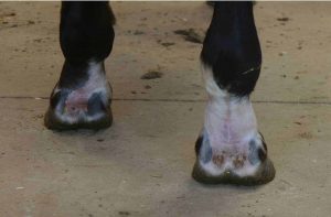
“This is particularly true for wounds affecting the distal (lower) limbs, a location with poor vascularization and oxygenation and little soft tissue,” she says. “Common complications for wounds in the distal limbs include extensive tissue loss and/or infection of the wounds, resulting in unrewarding treatment outcomes.
“The idea of using regenerative therapies is interesting because they have potential to simultaneously promote wound healing and reduce infectious burden,” she adds.
As summarized in the full-length article, in vitro (lab) research supports using peripheral-blood-derived MSCs and their secretome, consisting of bioactive factors MSCs secrete, in wound management.
“Mechanisms of action of the MSC secretome identified to date include promoting migration of dermal fibroblasts, promoting the formation of new blood vessels, and inhibiting the growth of various bacteria, including Escherichia coli and Staphylococcus aureus, even when present in biofilms,” Van de Walle says.
Harman adds that a handful of in vivo (in the live horse) studies exploring amnion, stem cells, and PRP have been published with variable yet overall positive results.
Results from one study support using equine amnion for bandaging certain distal wounds. In ponies, pinch grafts applied to full-thickness limb wounds were bandaged with amnion covered by an absorbent layer of gauze and elastic adhesive tape. Their median time to healing was significantly shorter (30 days) than that of a control group bandaged with nonadherent dressings covered with gauze and elastic adhesive wrap (39 days).
In another study researchers created wounds on nine horses and treated them with either amnion or live yeast covered with a nonadherent bandage. The rate of wound contraction and epithelialization did not differ between the groups; however, exuberant granulation tissue (aka proud flesh) severity was significantly less for amnion-treated wounds. It also took fewer days for wounds treated with amnion to completely heal.
Several research groups have explored MSCs in wound management.

“Injected stem cells appear more beneficial than topical application,” says Van de Walle. “This finding is based on a study that found significantly decreased wound areas when injecting umbilical-cord-blood-derived MSCs in the wound margins versus applying these cells topically embedded in a fibrin gel.”
Harman et al. also described a study using oral-mucosa-derived MSCs or their secretome mixed in a hyaluronic acid (HA) gel topically applied to surgically induced wounds on the thorax and forelimbs. Both HA mixtures (i.e., with MSCs or the secretome) had a positive effect on healing compared to the untreated control groups and the horses treated with HA alone.
Finally, PRP appears to hold great promise for promoting and supporting wound healing. In one of the many studies Harman et al. cite, they relayed data by Pereira et al. (2019), who evaluated various PRP preparations administered topically or injected into surgically created distal limb wounds.
“Key findings were that healing time was reduced in all PRP groups compared to controls, less granulation tissue was noted in the treatment groups, and the PRP gel had the most positive effects on wound healing,” says Van de Walle. “Overall, that study concluded that all of the studied PRP preparations positively affected healing and, thus, warrant further exploration.”
Conjunctival Stem Cells for Immune-Mediated Keratitis
Immune-mediated keratitis (IMMK) is a nonulcerative, nonpainful keratitis characterized by various degrees of corneal vascularization and opacification. The condition doesn’t typically lead to eye loss but might impair vision due to scarring, secondary infections, and diffuse corneal edema.
“IMMK is an increasingly diagnosed disease, which generally is well controlled by standard topical eye medication, such as cyclosporine,” says Brian Gilger, DVM, MSc, Dipl. ACVO, a professor at North Carolina State University’s College of Veterinary Medicine, in Raleigh. “But in many cases IMMK therapy either quits working or the IMMK is not responsive, leading to the need for new treatments.”
Therapy is typically long-term and, as many owners have learned firsthand, chronic ophthalmic therapy can be difficult in horses.
Because BM-MSCs modulate the immune system by downregulating inflammation, Gilger’s research team evaluated the efficacy of these cells for treating IMMK in a series of four horses (Davis et al., 2019).

Veterinarians administered autologous subconjunctival BM-MSC injections in all horses every three to four weeks for one to five treatments. Horses were maintained on a standard medical treatment regimen throughout the BM-MSC treatment period.
“These BM-MSCs are identical to those used in orthopedic applications,” Gilger notes. “And the injections are made with the horse lightly tranquilized, allowing the horse to return to normal activity within hours of the procedure.”
Three horses had increased corneal clarity, decreased neovascularization (new blood vessel growth, which can cause vision loss), and reduced surface irregularity. Only one of the treated horses was not responsive to subconjunctival BM-MSC therapy.
“These experimental results demonstrate the safety and potential efficacy of an innovative solution for IMMK,” says Gilger. “Subconjunctival autologous BM-MSCs can currently be used in clinical patients. Our current research is focused on determining if the cells themselves migrate to the area of inflammation or if they stay locally and secrete mediators to modulate the immune system. We are also examining the use of MSCs in other diseases, such as equine recurrent uveitis.”
Future Directions in Equine Regenerative Medicine
More than 25 years after equine veterinarians started using regenerative therapies, we are still scratching the surface of what they can do. Considering the power these therapies pack, they certainly warrant funding for additional well-designed and -controlled in vitro and in vivo studies. The many avenues that need to be explored include delivery methods and ways to produce the various therapeutic products. “Another exciting avenue of regenerative therapies is their potential as an adjunct or replacement for antibiotics,” says Van de Walle. Antibiotic overuse can lead to antimicrobial resistance—a potential health threat to humans and animals. The prospective of promoting tissue healing while also fighting bacterial infections is promising, particularly in wound management.”
Editor’s Note: This article originally appeared in the 2023 Research Roundup issue of The Horse: Your Guide to Equine Health Care.