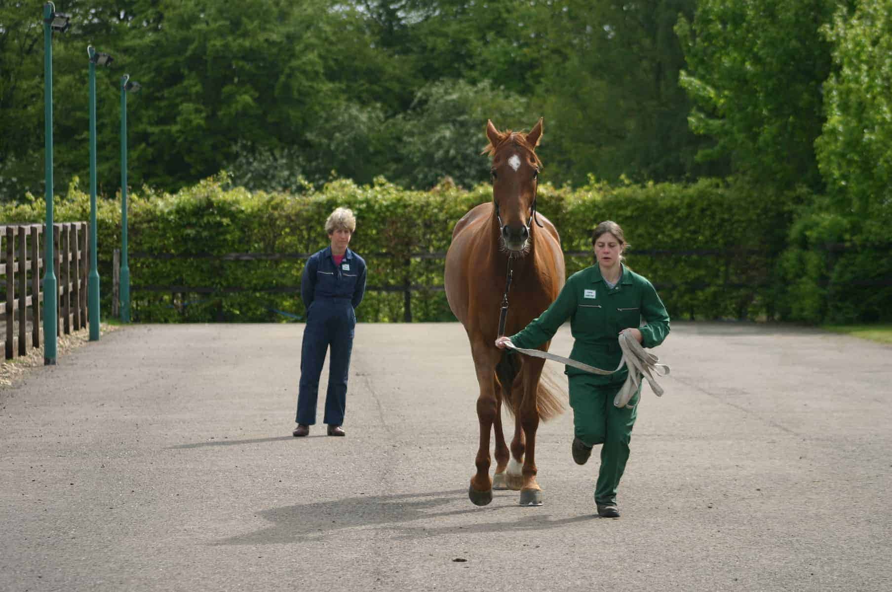
Equine Lameness Detection

Some veterinarians are adding a new tool to their lameness diagnosis arsenal
If ever there were a time we wish horses could speak our language, it’s when they are lame. If only Sir Runsalot could say, “You know, I was galloping around in the pasture today and I twisted my knee,” his owner could save time and money in veterinary exams and diagnostics to pinpoint the problem. But, unfortunately, Sir Runsalot does not speak our language, so his veterinarian interprets the situation to the best of his or her ability.
It takes veterinarians years of practice and evaluating countless horses to develop the keen eye needed to properly diagnose lameness. And even then some horses still baffle the experts. Fortunately, diagnostic methods such as digital radiography (X ray), magnetic resonance imaging (MRI), thermography, and scintigraphy (bone scan) developed in recent years can help veterinarians pinpoint exactly what is wrong. And the newest device on the block is a sensor system called the Lameness Locator. But nothing can or likely will ever replace a skilled professional’s discernment. All these diagnostic tools are just that–tools that help a veterinarian reach a probable conclusion and course of treatment.
Initial Examination
David W. Ramey, DVM, focuses on the care and treatment of sport and pleasure horses in his Southern California practice. In lameness exams he uses technology to complement his own comprehensive work-up and says such exams combine both objective and subjective analysis. For instance, the human eye is fallible and not always reliable in recording and processing information. Thus, two different veterinarians looking at the same horse might see two different legs as being lame.
When presented with a lame horse, Ramey first listens to the owner or trainer’s complaint and then begins evaluating the animal. “I can often get hints as to what the problem is from the owner or trainer, but I try not to get too focused too early,” he says. “After hearing what the owner or trainer has to say, I spend a good bit of time looking at the horse without watching it move–feeling, for example, for things like pulses in the feet, stiffness in joints, or swellings where swellings aren’t supposed to be. I learn a lot before I ever watch the horse move.” Based on these initial observations and upon watching the horse’s gaits, Ramey decides what other types of diagnostics are required.
Visual Interpretation
Lameness can begin gradually or acutely, and it can affect one or multiple limbs. Fortunately, horses are fairly predictable in the way they move when a leg hurts. Take a hind limb lameness, for instance: Veterinarians look for a “pelvic hike,” an upward movement of one side of the pelvis or a dropping or tilting of the pelvis to one side. However, this does not take into consideration the asymmetrical pattern of vertical movement of the entire pelvis during and after the stance phase (when the hoof is on the ground).
“The more objective description of pelvic movement with lameness allows one to see that sometimes it is less downward movement, sometimes it is a less upward movement of the pelvis, and sometimes it is both that are important for the detection of hind limb lameness,” says Kevin Keegan, DVM, MS, Dipl. ACVS, professor of equine surgery at the University of Missouri’s College of Veterinary Medicine. Then the veterinarian must consider head movement, which can be confusing depending on the specific lameness condition. While it is most commonly seen in front leg lameness, it can also occur in hind leg lameness.
When looking at head movement patterns, “the key observations in this sequence of events are the downward followed by upward head movement during stance, and upward followed by downward head movement during the swing phase of the stride,” Keegan reported in the American Association of Equine Practitioners (AAEP) 51st Annual Convention Proceedings (2005). “In the sound horse, the maximum and minimum vertical head positions during each one-half cycle of the stride are equal. The head never moves down to a lower position during stance phase of the lame limb compared with the sound limb, so if this is observed, the correct limb can always be identified. ‘Down on sound’ is generally a good way to observe forelimb lameness.”
Lameness Diagnosis
While the Lameness Locator is a helpful diagnostic tool, veterinarians employ a wide range of other lameness detection methods to pinpoint areas of concern, including:
- Flexion Tests Veterinarians regularly employ these subjective tests to exacerbate baseline lameness and reveal unknown internal issues in horses.
- Nerve blocks Veterinarians use analgesia to numb specific joints and tendon sheaths and block regional areas to isolate specific locations of pain.
- Radiographs (X rays) Veterinarians use these digital images to evaluate bone structures and detect issues such as arthritis and/or fractures.
- Magnetic resonance imaging (MRI) An MRI unit produces three-dimensional images of bone and soft tissue that can help veterinarians detect a wide range of abnormalities.
- Nuclear scintigraphy (bone scan) This imaging modality reveals “hot spots” of bone and muscle metabolism that can indicate remodeling due to stress, fractures, or other causes.
- Ultrasound This method is helpful for evaluating soft tissue structures by using sound waves to create a picture based on tissue density.
- Computed tomography (CT scan) This variation on X ray technology is particularly good for examining fractures and hard tissue (skeletal) problems.
- Thermography A trained practitioner uses a specialized camera to detect areas of increased or decreased infrared heat on a horse, which might indicate injury.
- Force plate/sensor technology Fixed plates in a ground surface or force-measuring sensors attached to feet or shoes can evaluate the dynamic forces on each leg to pinpoint potential problems or gait asymmetry.
- Video analysis This evaluation method might require markers on the horse as reference points and involves using accompanying software.
—Stephanie Ruff
The Lameness Locator
To help pinpoint the exact location of horses’ lameness, Keegan partnered with Frank Pai, PhD, professor of mechanical engineering at the University of Missouri, and Yoshiharu Yonezawa, PhD, at Hiroshima Institute of Technology, in Japan, to develop an inertial sensor system called the Lameness Locator. With this system, the operator attaches small sensors noninvasively to the horse’s poll behind the ears, on the right front leg midline directly above the hoof, and on the croup midline in front of the tail. Two accelerometers measure the up and down movement of the horse’s head and pelvis, and a gyroscope measures orientation in space of the right front leg. The system monitors and records these motions while the horse trots, and it transmits the data wirelessly to a portable handheld tablet PC. The operator then compares the data to information in the database from both sound and lame horses. Almost immediately, the computer analyzes the data and indicates whether the horse is lame on one or multiple limbs, and where. The report indicates the limb(s) affected, the severity of lameness within each limb, and when the horse’s pain is at its peak within the stride cycle. The entire procedure, from sensor application to report delivery, takes about 10 minutes.
Generally speaking, the Lameness Locator puts precise measurements to the visual observations veterinarians make. The human eye samples about 20 times per second, whereas the Lameness Locator samples 200 times per second. However, Keegan reiterated that this tool is merely an aid–it is not going to make an inexperienced person a lameness expert.
Ramey has found this tool to be helpful in several different types of situations. For example, an owner or trainer might be convinced a horse has a problem but it is not easily seen. “I’ve had several horses where it was obvious that there was no (lameness) problem with the horse, and the Lameness Locator helped convince people to be confident enough to approach their complaint from a riding and training perspective, as opposed to thinking that the horse needed some sort of medication or injection,” he says.
Ramey has also used this tool in prepurchase exams, as another way to show that a horse is sound at the time of examination. He’s even had requests to collect baseline data on some horses simply as a record of how the horses travel normally.
“It does what we all try to do–look at the movement of parts of the horse’s body–it just does it with a great deal more sensitivity than the human eye,” he adds.
Ramey and Keegan agree that the Lameness Locator is not a blanket solution for lameness diagnosis. “There’s no cookbook approach to lameness diagnosis,” says Ramey. “Some problems can be solved with just a thorough physical examination.
“Sometimes figuring out the source of lameness may require diagnostic anesthesia; if I can make the area that hurts numb, the horse will begin to travel sound, and I can focus my efforts on that particular area,” he continues. “X rays (plain film, digital, and computed radiography) all have their place, and ultrasound are standard tools. In selected cases, scintigraphy and MRI might be used.”
The Next Steps
Keegan says the biggest hurdle with this new tool is educating veterinarians. In August 2011 the Lameness Locator’s manufacturer began its National Demonstration Program to equine veterinarians. Today there are about 75 practices using the technology, and they range from solo-veterinarian mobile practices to large clinics to universities to international clients.
Currently, in a research project funded by a National Science Foundation grant, Keegan et al. are working to develop another set of algorithms for evaluating the cantering horse. They are also looking at trot data collected previously and comparing it with the eventual diagnoses to try to equate patterns of movement to specific lamenesses. Because a problem could be caused by issues in more than one area, the research team is developing ways to help veterinarians detect and diagnose multiple limb lameness. They are also collecting and analyzing data on racking and pacing gaits. However, Keegan says that the rack is a difficult gait to evaluate, as the horses don’t maintain it for very long (and, besides, veterinarians don’t typically use it to evaluate lameness).
More To Come
Keegan hopes more veterinarians will learn to use the Lameness Locator to help them accurately diagnose horses–a trend Duncan Peters, DVM, of Hagyard Equine Medical Institute’s sport horse division, in Lexington, Ky., sees forming: “I think there will be an increase in the use of these technologies. It is driven by the desire to have an objective way to analyze problems. It will never replace the ‘art of lameness evaluation’ but will be very important supplementally to help diagnose and then monitor problems through rehabilitation.”
Written by:
Stephanie Ruff, MS
Related Articles
Stay on top of the most recent Horse Health news with


