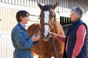
The equine fetlock is an amazingly complex, fist-sized structure that supports heavy loads. This is particularly true in racehorses running frequently at high speeds, making it a common site of injury in this horse population.
As part of the Welfare and Safety of the Racehorse Summit webinar series, Katie Garrett, DVM, Dipl. ACVS-LA, from Rood & Riddle Equine Hospital in Lexington, Kentucky, described how veterinarians use imaging modalities to diagnose fetlock issues and the common injuries they might find.
Fetlock Anatomy
Garrett emphasized that much of understanding diagnostic imaging is knowing your anatomy—she briefly reviewed the structures within the fetlock joint.
The four major bones are the cannon bone (MCIII, or third metacarpal), a pair of proximal sesamoid bones, and the long pastern bone (P1). Many soft tissues overlie those bones, “but we’re going to focus on the suspensory ligament branch (the suspensory ligament starts right below the knee and inserts onto the top part of the proximal sesamoid bones) and the distal sesamoidean ligaments, which originate at the bottom of the proximal sesamoid bones and extend down into the pastern region,” said Garrett.
Articular cartilage lines the surface of the bones within the joint. In healthy joints, that cartilage acts as a protective layer, said Garrett, preventing the bones from contacting each other directly. The bone just below the cartilage is the subchondral bone.
Diagnostic Imaging Options
The goal of imaging is to get an accurate diagnosis, after which the practitioner can create an appropriate, targeted treatment for the specific problem. Garrett described the six major imaging modalities veterinarians typically use to diagnose fetlock injuries—radiography, ultrasound, MRI, CT scan, nuclear scintigraphy, and PET—and their strengths and weaknesses. The first four are structural, meaning they provide veterinarians with information about the bone’s makeup:
- Radiographs provide good general images of bones, said Garrett. They’re relatively inexpensive (on average, a few hundred dollars for a set of fetlock radiographs), and veterinarians can acquire images at the barn. “Especially with the advent of digital radiography, oftentimes we can make a stallside diagnosis,” she said. “Some of the challenges associated with radiography are that it’s not as sensitive as CT or MRI for bony problems, and it gives us limited soft tissue information.” Overall, she said, it’s a good initial choice for assessing bony problems.
- Ultrasound provides good information about soft tissues and bony margins. Like radiography, it’s inexpensive and can be performed at the barn. “It’s not as sensitive as MRI for soft tissue problems, and it can’t image deep to bone or air, so we can’t see what’s going on inside a bone,” said Garrett. But for most soft tissue problems, it’s a good initial choice.
- MRI is a cross-sectional imaging modality, allowing veterinarians to get high-detail images because they’re looking at very thin slices of tissue. “It gives us superior info about tendons, ligaments, cartilage, bone bruising, and other bone damage,” Garrett said. “In the horse, we can only image the limbs from the knee or hock down. (Horses) do need to come to a hospital, and some systems require anesthesia.” Overall, MRI is an excellent choice for evaluating soft tissue and bony injuries, she said.
- Computed tomography (CT) is another cross-sectional imaging modality, but it excels at producing images of bones versus soft tissues. Garrett said it has a reconstruction ability that’s helpful when looking at complex fractures. Like when using MRI, however, veterinarians are limited to the portion of the limb they can image, horses must come to the clinic for imaging, and some systems require anesthesia. “CT is a very great choice for bony injury,” she said.
Garrett then described the two functional imaging modalities that provide veterinarians with information about what the bone is doing:
- Nuclear scintigraphy reveals information about bony turnover – where in the skeleton is there increased bone turnover activity. “Because we are able to image the entire skeleton in one session (it takes a few hours but is feasible), it’s a really good screening tool for bone problems in a lot of places in the skeleton,” said Garrett. “It’s relatively low resolution so typically requires an additional structural imaging modality such as radiographs for a more precise diagnosis.” Nuclear scintigraphy provides limited information about soft tissues and must be performed at a clinic. Overall, she said, it’s an excellent choice for stress fractures or other injuries that involve bony turnover.
- PET scan, a relative newcomer to the equine imaging scene, also evaluates bony turnover but provides much higher resolution and cross-sectional images, allowing veterinarians to pinpoint problems more precisely, said Garrett. It can only image from the knee or hock down, and horses do need to come to a clinic, but one recently developed scanner does not require general anesthesia. “We don’t have as much information on PET scanning as we do these other modalities because it is much newer,” she said, “but there’s a lot of really interesting work happening, and hopefully this going to be a really great tool in our toolbox.”
There’s no such thing as an ideal imaging modality, said Garrett. “If I could design it though, an ideal imaging modality would be inexpensive, very quick to acquire (minutes), we’d be able to perform it at the barn, it wouldn’t require general anesthesia, and it would be highly accurate,” she said.
“None of them can do everything, but if we look at radiography and ultrasound, they shine in the convenience and expense categories,” she continued. “And to be completely honest, most of the time we can make a diagnosis using radiographs or ultrasound. There’s a reason they’re so common; they’re generally very good. But sometimes we do need to move to our more advanced imaging modalities.”
Those modalities (MRI, CT scan, nuclear scintigraphy, and PET) generally cost more and require a hospital visit. “The tradeoff is you’re going to ideally get the answer you need,” said Garrett.
No matter the imaging modality used, Garrett emphasized the importance of interpreting results in light of the clinical examination. “We are never just treating images, we are always treating an actual horse,” she said. “Just because we find an abnormality on an imaging study doesn’t mean it’s significant. One of the difficult questions to answer is which abnormalities are significant to this particular horse. So that’s where we bring in our clinical examination in combination with our diagnostic imaging—they’re really going hand in hand.”
Take-Home Message
Due to the variation between horses and the overlap between changes seen in bone’s normal response to training and those seen in a pre-injury state, it’s currently impossible to use diagnostic imaging to predict lameness and determine whether a horse needs a break before injury occurs, said Garrett.
You could have two sets of images that look nearly identical, yet one could belong to a horse that’s lame while the other belongs to a sound horse that goes on to win a stakes race.
“That’s part of why we need to treat the actual horse and not the images,” she said. “There’s no magic machine that spits out the answer.”