Common causes of lameness in the hind end and how to diagnose and treat them

The general public might assume that a 1,200-pound horse is a pretty robust creature. Horse people, however, have learned from experience that this is not necessarily true. At risk of suffering wounds, sickness, or lameness, horses can be surprisingly fragile animals. Anyone who has ever owned a horse has likely dealt with subtle and frustrating lamenesses. While forelimb lamenesses are fairly common, hind-limb issues can be less obvious and even underdiagnosed.
Identifying the Issue
What exactly does a hind-limb lameness look like? Many owners are familiar with front-end issues, which are often easier to detect with the naked eye. A head bob, or a dramatic raising of the head when the lame leg bears weight, is a telltale sign of forelimb lameness. But what hind-limb indicators does your veterinarian look for during a lameness exam?
Lameness in the hind end can present in a couple of ways. When I’m doing a lameness work-up, I like to incorporate both longeing on a circle and trotting in a straight line to try to determine where the lameness appears. Sometimes having a rider work the horse under saddle can accentuate subtle issues. Excessive hip movement or “hip hikes,” an unwillingness to bring a leg forward, toe dragging, and a “bunny hopping” canter are just a few routine signs of hind-end lameness. No two presentations are the same.
Imaging the Hind Limb
Before choosing an imaging method, veterinarians perform diagnostic analgesia (joint blocking) to isolate lameness to a certain area. Because vets perform most of their equine work on the farm, stallside radiographs (X rays) are the modality of choice for evaluating any bone or joint in this setting. Radiography, however, does not reveal many details about soft tissue. Ultrasound is ideal for evaluating tendons, ligaments, or other soft tissue structures in the field.
Your veterinarian might choose to perform advanced imaging back at the hospital. There, he or she can use nuclear scintigraphy to better pinpoint a problem area, along with magnetic resonance imaging (MRI) or computed tomography (CT) to further evaluate more insidious conditions such as proximal suspensory ligament desmitis, navicular bursitis, or sesamoidean ligament conditions. Let’s review five conditions they might find.
Osteoarthritis (OA)
Osteoarthritis is not just your grandmother’s disease—it’s also a common equine condition. While it’s most common in geriatric horses, OA can have devastating effects on any equine athlete, dramatically interfering with performance.

“OA starts as inflammation in a joint (synovitis), progresses to arthritis when the cartilage becomes involved, and finally to osteoarthritis when the bone surrounding the joint starts to remodel,” says Matthew G. Durham, DVM, Dipl. ACVSMR, of Steinbeck Peninsula Equine Clinics, in Salinas, California.
Cartilage has incredible compression and tensile strength. Synovial fluid, which is produced by the lining of the joint sac, acts as an outstanding lubricant for joint surfaces and also provides nourishment for cartilage. “When the joint becomes inflamed the lubricating … abilities are greatly decreased, thereby magnifying the traumatic forces on the cartilage and bone,” says Durham.
While OA can develop in any joint, it’s a common cause of hock pain. The hock comprises four joints. The lowest two, the distal intertarsal and tarsometatarsal joints, are especially susceptible.
“With distal hock OA, it is common for both limbs to be affected, which means the clinical signs can be a little less obvious and can creep up over time,” says Durham. “In horses where hard stops are common, such as in many Western disciplines, the horse may tend to overuse the front end when stopping or have a slower, stuttering stop.”
Thankfully, veterinarians have many OA management and treatment options available to them. The least invasive products are nutraceuticals, better known as oral joint health supplements, containing glucosamine, hyaluronic acid (HA), and other compounds.
Practitioner-prescribed non-steroidal anti-inflammatories (NSAIDs) such as phenylbutazone and firocoxib can also be helpful. However, chronic usage does increase a horse’s risk of developing gastric ulcers and kidney disease, so discuss these drugs with your veterinarian.
“Systemic injectable treatments are often effective,” Durham says. “Legend (intravenously administered HA) helps to restore synovial fluid to its normal state and is picked up by inflamed joints anywhere in the body. Adequan (intramuscularly administered polysulfated glycosaminoglycan) is incorporated more directly into cartilage. In horses with multiple joints affected, particularly geriatric horses, these treatments can be very effective.”
Hock injections are one of the most common joint injections veterinarians perform in the field. But not all injection cocktails are equal. Of the many combinations available, corticosteroids are the mainstay. They work to reduce the inflammation in OA cases and minimize damage to synovium and cartilage.
“Newer joint injection treatments leverage the body’s own immune system to treat itself,” says Durham. “Various systems use different tactics but, in general, blood is drawn and processed in a way that encourages expression of certain proteins which have potent anti-inflammatory effects and encourage healing, or for harvesting platelets.”
Platelet-rich plasma (PRP) and IRAP (interleukin-1 receptor antagonist protein) are common biologic products used in joint injections that replicate these features. Durham says polyacrylamide gels show promise in the future of equine joint injections. These devices “mimic the physical qualities of synovial fluid and stay present in the joint.”
Osteochondrosis Dissecans (OCD)
Osteochondrosis dissecans is a disease that mostly affects younger horses. Caretakers often recognize it just as horses are starting under saddle. The horse can be progressing well, then suddenly comes up lame with a swollen stifle, for instance.
“The bone and the cartilage in joints with OCD don’t form normally,” says Steve Trostle, DVM, Dipl. ACVS, ACVSMR, co-owner of Blue Ridge Equine Clinic, near Charlottesville, Virginia. “Subsequently, the cartilage and bone underneath it become irregular in thickness and weaker than in normal joints. This can lead to the formation of cartilage and bone flaps that can remain partially attached to the bone or break off and float around in the joint. Loose flaps and areas of abnormal cartilage and bone cause inflammation in the joint and over time may lead to the development of arthritis.”
We don’t fully understand the exact cause of OCD. While genetics play a role, researchers are working to understand other potentially influential factors. One key area of study is nutrition, because many suspect excess energy intake contributes to OCD lesions.
The disease can occur in any equine joint but most often manifests in the fetlocks, hocks, and stifles. While owners can pursue conservative therapies such as oral medications and joint injections, surgery remains the gold standard in most cases. Today the most common way surgeons address OCD lesions is with arthroscopy, where he or she passes a small camera into the joint to view the lesion, then inserts surgical instruments to remove it.
“Arthroscopic removal is typically performed for smaller to moderate-sized fragments and generally has a very good prognosis,” says Trostle. “The more difficult OCD conditions are those that are large and on direct weight-bearing surfaces. In those cases some (surgeons) have moved away from removal and debridement and are now trying to secure the OCD flap with pins and augmenting with (injectable) biological therapies.”
Sacroiliac (SI) Joint Disease
More and more veterinarians are recognizing sacroiliac—the area where the spinal column meets the pelvis—pain as a cause of hind-limb discomfort, particularly in performance horses. Common signs of pain in this region include a reluctance to go forward, lack of impulsion, and an uncoordinated “bunny hop” canter, among others.
“The sacrum is a section of five fused vertebrae located between the lumbar spine and coccygeal (tail) segments,” explains Luvie Abell, DVM, of Grand Prix Equine, a sports medicine practice in Hawleyville, Connecticut. “Its widest point, known as the sacrum, connects to the underside of the pelvis, known as the ilium. This connection forms the sacroiliac joint.
“Subluxation (misalignment) and osteoarthritis are the main causes of discomfort to the sacroiliac joint,” she continues. “This may be exacerbated by a more pressing source of pain,” often pointing to problems in the hock or stifle that force the SI to compensate.
Because the SI joint is beneath inches of muscle, imaging the area can be challenging. “This region is far too large to simply shoot a radiograph the way that we can with a hock or stifle,” says Abell. “So, we are left primarily with ultrasound and nuclear scintigraphy (bone scan).”
Treatment options exist but can be limited. “If your veterinarian has diagnosed a source of pain specifically to the sacroiliac joint, you will likely be discussing corticosteroid injections and chiropractic adjustments” as treatment options, she says.
If these therapies resolve the lameness (and you’ve addressed any other sources of hind-limb discomfort that might be exacerbating SI pain), Abell says it’s important to maintain the horse’s comfort with a good fitness regimen that allows the gluteal muscles to develop and properly support this area of weakness.
Upward Fixation of the Patella
Commonly known as a “weak stifle,” upward fixation of the patella (UFP) is a relatively prevalent condition in which a horse’s hind leg gets stuck in extension. UFP can manifest in young horses in early training, but it can also be associated with straight hind-limb conformation or lack of proper musculature in all ages of horse.
“A horse’s inability to release the passive stay apparatus (a network of muscles, tendons, and ligaments that allows a horse to stand with minimal effort) of the stifle results in upward fixation of the patella,” says Michael Caruso III, VMD, Dipl. ACVS, owner of Reedsdale Equine Specialists of Central Tennessee.

Treatment options range from conservative to very radical. “The easiest treatment option involves alterations in your horse’s training to increase the development of the quadriceps muscle,” says Caruso. “Increasing this muscle’s strength will effectively ‘tighten’ the patellar ligaments, thereby correcting upward fixation of the patella in some horses.”
You can help strengthen these muscles by trotting for extended periods, doing hill work, and working in deep footing. Caruso advises owners against cantering these horses on the longe, however, which can contribute to patella trauma.
If exercise changes fail, surgical options include medial patellar ligament splitting, which involves making several small incisions in the medial patellar ligament. “This form of splitting has been shown to increase the ligament diameter by two- to threefold over four to six weeks,” in hopes the stifle will stabilize, says Caruso.
Soft Tissue Injuries
Soft tissue strains and tears and resulting lameness are all too common. Ones veterinarians frequently diagnose in hind ends include tendinitis, suspensory ligament injuries, and degenerative suspensory ligament desmitis.
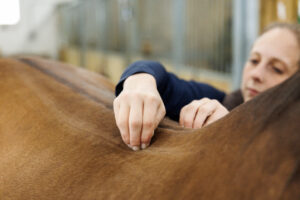
When first examining a soft tissue injury on the farm, your veterinarian will likely ultrasound the area of suspicion.
“Once you can establish the amount of damage, then you can determine what that horse can do,” says Grant Myhre, DVM, owner of Myhre Equine Clinic, in Rochester, New Hampshire.
Rest is certainly a mainstay in treatment, but it is not a cure-all. “Controlled exercise is really important,” says Myhre. “You want there to be function, you want to have enough exercise to enhance the rehabilitation of the soft tissue structures.”
Bringing a horse back from soft tissue injury involves returning him to work slowly, either by hand- or tack-walking. Myhre stresses that bringing a horse back to work too quickly can be catastrophic. “Once you overdo it, you start breaking down tendon or ligament fibers,” he says.
Rehabbing a soft tissue injury can be a trial-and-error process. “Is the lameness getting better? Is the tendon hot or still inflamed? If so, we may have to decrease exercise,” says Myhre.
Your veterinarian might recommend extracorporeal shock wave therapy, which involves applying acoustic wave pulses to soft tissues to improve healing. He or she might also suggest injecting biologic products such as PRP, IRAP, or stem cells into core lesions to speed the healing process. Surgical approaches, such as desmotomy (tendon splitting) and neurectomy (cutting the nerve), might even be beneficial depending on the condition. Proper imaging is pertinent to guide your horse’s treatment protocol and give him the best opportunity for a successful recovery.
Take-Home Message
Hind-limb lameness can plague performance and backyard horses alike. Whether the condition is sudden and acute or insidious and chronic, you have the best chances of diagnosing and treating it with your veterinary team’s guidance.