5 Points to Help Veterinarians Diagnose EPM
Equine protozoal myeloencephalitis remains challenging to definitively diagnose. Here, a practitioner offers an update about the disease.

Equine protozoal myeloencephalitis remains challenging to definitively diagnose. Here, a practitioner offers an update about the disease.

A high-quality and well-set-up ultrasound image can mean the difference between an accurate or missed diagnosis in equine practice.

Equine biosecurity might become easier thanks to new in-field tests using recently developed isothermal technology, which could detect infected horses in less than an hour.

Use this visual guide to learn about the technologies your veterinarian might use to look inside your horse and why.
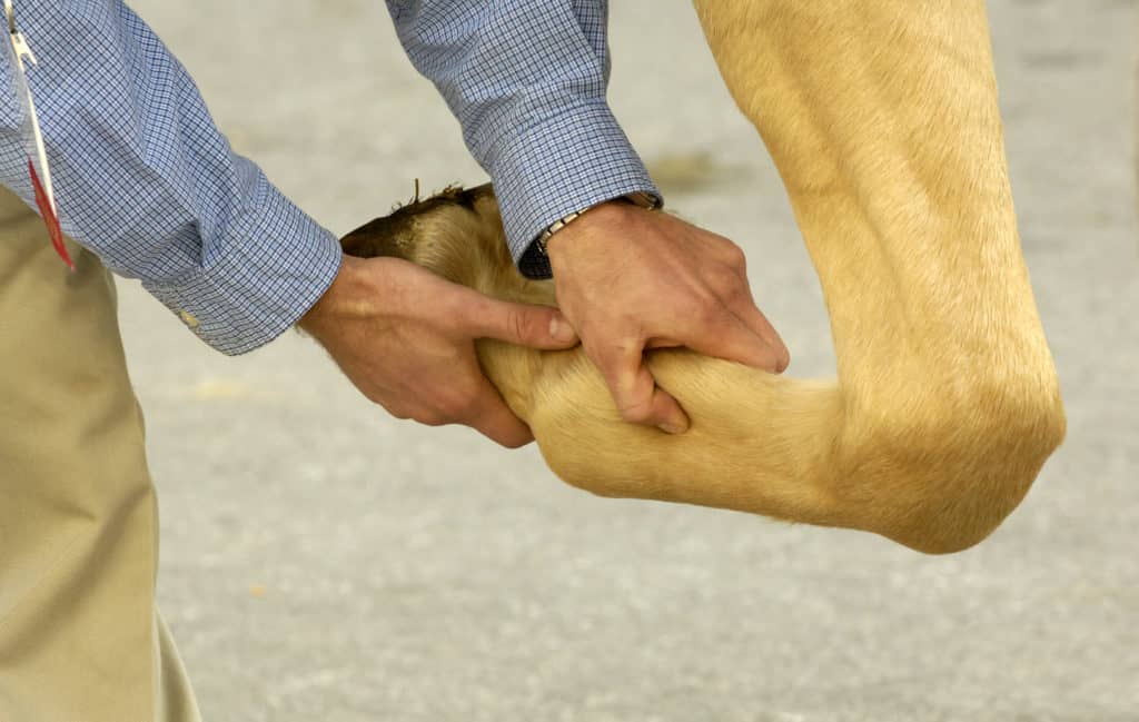
The editors of The Horse: Your Guide To Equine Health Care have scoured our archives and gathered our top lower limb lameness content. Sponsored by Hallmarq Veterinary Imaging.

Poll pain can cause performance, behavior, and welfare issues for horses. Learn how vets diagnose and treat it.

In the four months since the MILEPET scanner’s installation at UC Davis Veterinary Hospital, veterinarians have imaged 100 horses; more than half were performance and pleasure horses.

Learn from Dr. Jennifer Janes, part of the University of Kentucky’s CSI team for horse diseases, conditions, and poisonings.

He might seem perfect—but before you call him yours, determine if a horse is sound and serviceable for the job at hand and if you can live with his inevitable flaws.

Read about three real-life examples of equine athletes that made full recoveries from their injuries, including their diagnostic challenges, rehab modalities, and recovery details.

Here’s what to expect if your horse needs to undergo gastroscopy, the only surefire way to check for equine gastric ulcers.

Researchers described normal pituitary gland appearance on MRI. Their findings might help veterinarians identify PPID in horses and start treatment earlier.
Learn what steps you and your veterinarian can take to get to the bottom of subtle horse health problems.

Computed tomography (CT) and magnetic resonance imaging (MRI) are two diagnostic imaging methods veterinarians can use to capture images of structures within your horse’s body. Learn more in this visual guide!

Veterinarians could soon determine which horses are at risk of certain neurologic diseases through a simple urine test that reveals how a horse breaks down vitamin E.

Researchers studied these rare mineral concretions, how to best detect them, and commonly found concurrent conditions in affected horses.