How veterinarians diagnose pathology in these difficult-to-visualize regions of the horse’s body
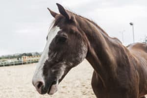
It started with a sour attitude while tacking up, then progressed to stiffness under saddle, difficulty making transitions, and a poor-quality canter. Now your horse is throwing in uncharacteristic bucks during rides. He might not be simply misbehaving; he might be telling you his neck or back hurts.
While we typically think of limb lameness as being the main reason for performance limitations in horses, problems within the spine can also cause reduced athleticism, changes in behavior, altered movement, weakness, neurologic signs, and even lameness. To investigate a suspicion of pathology (disease or damage) originating from this area, veterinarians often pursue diagnostic imaging of these regions. The challenge is large groups of stabilizing muscles that can impede visualization surround these structures. So, what are our options for imaging them? In this article I’ll describe the modalities veterinarians use, based on case studies designed to exemplify those we often see in the field.
A Dressage Horse With Neurologic Deficits
Let’s begin with the neck. The first case is a 5-year-old Warmblood mare that has just begun dressage training. Her owner noticed she trips occasionally under saddle, particularly when working in a frame, but has noted no other performance issues. The mare’s veterinarian conducts a physical examination and notices some neurologic deficits, which are behaviors or movements that indicate abnormal spinal cord or nerve function. These can include signs like loss of balance and the horse stepping on herself, dragging her toes, or swinging her limbs wide during the neurologic examination. The deficits the veterinarian in this case noted indicated a possible problem in the mare’s neck.
In these types of cases the first imaging study veterinarians invariably perform is radiography (X rays) of the horse’s neck, which must remain straight with the head still. Therefore, horses are typically sedated for this study. In some cases X rays might reveal an obvious abnormality, such as narrowing of the canal within the vertebrae through which the spinal cord runs. However, it is not uncommon for these horses to have normal X rays, despite the veterinarian’s strong suspicion of neck pathology.
In this instance the practitioner might recommend more advanced imaging to determine if the horse’s spinal cord, or the nerves exiting it, are being compressed. One way to do this is using a technique known as myelography. A myelogram involves placing the horse under general anesthesia, injecting a dyelike substance into the fluid surrounding the spinal cord, and repeating neck X rays, usually while also manipulating the horse’s neck into varying degrees of flexion and extension. The injected dye shows up as bright white on X rays and surrounds the spinal cord. If the layer of white fluid surrounding the cord is narrowed or interrupted, it might indicate compression of the spinal cord in focal areas, leading to the horse’s neurologic signs.
The final study vets might perform in these (and osteoarthritis) cases is computed tomography (CT) of the neck. They often perform this three-dimensional imaging study in conjunction with myelography. A neck CT enables vets to check for narrowing of the spaces through which the nerves exit the spinal cord that might be contributing to the horse’s clinical signs, among other issues. Combined CT/myelography is the most comprehensive way to assess a horse with neurologic signs localized to her neck.
A Barrel Racer With Osteoarthritis
The second case is a 12-year-old Paint Horse gelding used for barrel racing. His owner reports reluctance to bend to the right, and the veterinarian notices neck stiffness but no neurologic abnormalities. Again, the attending veterinarian is likely to begin with neck X rays under sedation, this time to assess for issues such as osteoarthritis, which might be causing neck stiffness without neurologic deficits. He or she might follow this examination with an ultrasound exam of the neck. Practitioners do not typically perform this imaging modality in horses with neurologic abnormalities because the vertebrae block the ultrasound waves, obscuring the view of the spinal cord. In horses with osteoarthritis, however, ultrasound can allow veterinarians to assess the severity of degenerative changes and can also guide injections of therapeutics such as corticosteroids or biologics into the arthritic joints to improve the horse’s comfort. Ultrasound exams and, particularly, ultrasound-guided injections also require the use of sedation to keep the horse still and with his head hanging low enough to enable the veterinarian to reach the areas of interest. Practitioners rarely need advanced imaging such as CT to diagnose and manage cases such as these.
Focusing on the Back
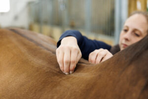
Moving onto the back, indications for imaging this region of the horse might include unusual behavior under saddle, such as reluctance to work, a restricted or altered way of going, head-tossing, and bucking. The owner might have also noticed the horse resents saddling. On clinical exam the horse might exhibit signs of discomfort or pain when the veterinarian touches, palpates, or otherwise manipulates the animal’s back. The decision to initially pursue X rays, ultrasound, or nuclear scintigraphy (aka bone scan) to investigate these clinical signs might depend on several factors.
First: Which region of the back does the veterinarian suspect is the problem? The thoracic spine (withers to just before the lumbar area) can be better seen with X rays than ultrasound, particularly because the adjacent air-filled lungs are easier for an X ray beam to pass through than the thick musculature of the horse’s lumbar spine, (between where the last rib connects and the sacrum). In contrast, ultrasound might yield more valuable information in the lumbar spine. It is worth noting that some lower-power, portable X ray machines struggle to penetrate well even in the thoracic spine. For this reason veterinarians might recommend transporting the horse to a local clinic with a more powerful in-house machine to obtain the best diagnostic images.
Again, the veterinarian will likely perform both X ray and ultrasound studies with the horse under sedation to limit motion. The most common abnormal findings on imaging the back are osteoarthritis and spinous process impingement (aka kissing spines). Both high-power X rays and ultrasound can help practitioners detect many of these back abnormalities, although they generally can see lumbar spine osteoarthritis best on ultrasound and kissing spines most optimally on X rays.
Kissing spines has become a hot topic within the equine community. Veterinarians use the term when the normal spaces between the bony extensions that project upward from each of the back vertebrae (spinous processes) narrow or disappear, causing the processes to touch one another. The problem with using radiographs to detect this pathology is evidence of these changes on X rays does not always represent a source of pain for the horse. For example, as reported in a 2004 study by Erichsen et al., veterinarians have noted narrowed spaces between the spinous processes on X rays in horses with no signs of back pain that are happily competing in a variety of disciplines. Therefore, if X rays show signs of kissing spines but the horse is showing nonspecific signs such as altered movement without specific evidence of back pain, the veterinarian might recommend additional imaging to confirm these changes as the cause of clinical signs (as opposed to a leg lameness, for instance).
The next logical imaging option is nuclear scintigraphy, which involves injecting the horse with a radioactive substance that attaches to regions of active bone remodeling. For this reason it can indicate whether the changes seen on the X rays are associated with an active process within the bone that’s likely to be a source of pain for the horse.

Take-Home Message
As you can see, a horse’s specific clinical signs often dictate exactly how veterinarians plans their approaches with regard to diagnostic imaging of the neck and/or back. They must select imaging studies carefully to get the most pertinent information to guide treatment recommendations on a case-by-case basis. These advanced forms of evaluation, however, are not available everywhere, can be expensive to pursue, and often require referral to a clinic that offers them.