Is My Horse Neurologic or Lame?

In the something-else-altogether category lives a sizable list of neurologic conditions that can be tricky to spot early and difficult for a veterinarian to diagnose. Further complicating things, neurologic issues might first be apparent as signs other than an altered gait.
Therefore, it’s important that horse owners and caretakers understand the signs of neurologic disease and keep an eye out for them just as they do indications of gastrointestinal or infectious disease. And as soon as clinical signs or unusual behavior appear, it’s crucial they call a veterinarian.
Two equine practitioners with vast neurology experience shared with us what neurologic signs owners might see, what practitioners look for during lameness and neurologic exams, and how they diagnose.
Lameness, or an abnormal stance or gait caused by either a structural or a functional disorder of the locomotor system, is a clinical sign, not a disease. Neurologic lameness, generally seen as ataxia, or incoordination, can be caused by bacterial, viral, protozoal, or rickettsial infections; trauma or congenital or developmental diseases; degenerative diseases or cancers affecting the brain or spinal cord; and toxicities.
Equine protozoal myeloencephalitis (EPM) is the most common infectious cause of neurologic lameness. Rarer infectious causes include tetanus, botulism, Lyme disease, rabies, West Nile virus, equine encephalomyelitis, and equine herpesvirus-1.
Dr. Stephen Reed
Musculoskeletal vs. Neurologic
It can be difficult to detect whether a horse’s lameness is musculoskeletal or ataxic, says Stephen Reed, DVM, Dipl. ACVIM, of Rood & Riddle Equine Hospital, in Lexington, Kentucky. Reed is a recognized authority on equine neurology who has spoken extensively on wobbler syndrome, EPM, and head and spinal cord trauma, as well as peripheral neuropathy, all of which can cause neurologic gait deficits.
He says differentiating between the two is challenging because deficits can be similar and distinguishing features subtle. “The often simple sentence used to describe the difference between lameness and ataxia is that ‘A lame horse is regularly irregular, and an ataxic horse is irregularly irregular,’ ” says Reed. “The deficits one looks for in an ataxic horse are weakness, ataxia, dysmetria, and spasticity.

Amy Johnson, DVM, Dipl. ACVIM, assistant professor of large animal medicine and neurology at the University of Pennsylvania’s New Bolton Center, in Kennett Square, also looks for the irregularly irregular or regularly irregular distinction.
“Musculoskeletal lamenesses tend to be predictable (happening with every step), and neurologic lamenesses are more likely to be unpredictable,” she says. “It is not unusual to see a horse that has both problems, neurologic disease and musculoskeletal disease. Those horses can be even trickier to figure out.”
Johnson adds that one clinical sign that indicates a lameness is neurologic in nature is neurogenic atrophy, which is severe muscle wasting that has developed quickly due to a loss in neural stimulation that helps maintain muscle fiber mass.
Additional clinical signs owners might notice include an abnormal stance, proprioceptive (awareness of one’s limbs) problems, paralysis, muscle twitching or spasms, falling, and problems lying down and/or getting back up.
Despite these red flags, owners still sometimes think they’re dealing with a simple musculoskeletal lameness and put off calling the vet. “A veterinarian should be called whenever the owner becomes concerned, if it worsens or doesn’t improve quickly, or if there is no obvious explanation for the lameness,” says Johnson.
Because certain neurologic conditions can progress quickly, it is important to get a veterinarian involved as soon as signs appear so the horse has the best chance at recovery. Early intervention can also cost less in the long run.
The Neurologic Exam
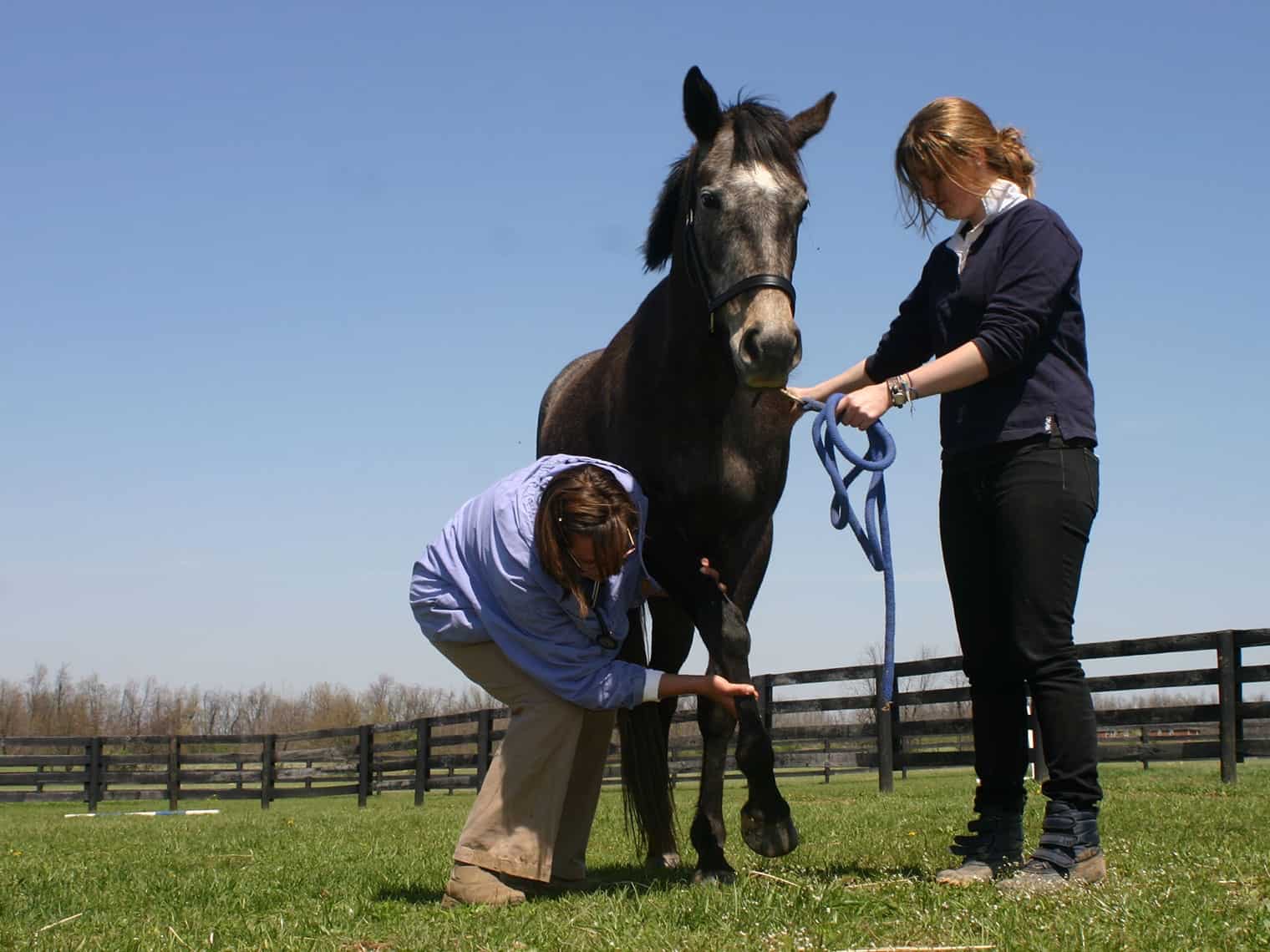
Reed says the physical exam is key to distinguishing whether a lameness is neurologic or musculoskeletal. Anytime a veterinarian needs to examine a horse, he or she first gets a history, asking the owner what the horse is like when healthy and what changes he or she has noticed, such as if the horse is urinating and defecating normally. The veterinarian also records the horse’s vital signs and evaluates overall health.
Then it is on to the neurologic exam, which Reed begins at the head and proceeds toward the rear.
The exam includes the following steps, as described by Reed and Johnson, although the order might vary among veterinarians:
- The veterinarian examines the horse’s attitude, behavior, and mental status/state of consciousness, looking for signs of dullness, lethargy, stupor, or unresponsiveness. He or she might present different stimuli to see how the horse reacts. For instance, normal horses should be bright, alert, and responsive and blink if someone waves a hand in front of their eyes.
- The vet then performs a detailed cranial nerve exam to evaluate the 12 nerves that supply the head structures. With these tests he or she assesses all motor (muscle) and sensory functions, including of the eyes, ears, muzzle, jaw, and tongue. The veterinarian checks the size of the pupils and whether they respond to light. He or she looks at skin and muscle sensation on the face, cheek, and up the nose. In addition, he or she watches how the horse swallows.
- The practitioner evaluates the horse’s posture, stance, and musculature. He or she palpates muscles and joints looking for any pain reaction, asymmetry, lack of muscle tone, muscle wasting, swelling, heat, and unusual lumps. The vet might also test the horse’s range of motion through flexion and extension of the joints and by moving the horse’s neck.
- Then the veterinarian tests reflexes by stimulating an area of skin with a ballpoint pen or a similar object and looking for an appropriate muscle reaction. For example, during the cervicofacial reflex, the veterinarian stimulates the neck and observes the face for appropriate ear twitching and lip grimacing.;
- The practitioner tests the horse’s proprioception by placing his feet in unusual positions and observing how the horse places his limbs during ­movement.
- Finally, he or she examines the horse’s gait looking for irregularities, head-bobbing, head-tilting or -shaking, toe-dragging, or incorrect placement of limbs during movement. The veterinarian has the horse walk and trot in a straight line, including over different surfaces. He or she also has the horse walk in circles, a zig-zag, and in a serpentine; with his head and neck elevated; while the tail is pulled; over a curb; and up and down hills with the head in a normal as well as an elevated position. If a horse is ataxic, the veterinarian might try to determine if the horse is hypermetric (has a long-strided, spastic gait), hypometric (stiff or spastic movement with limited joint flexion), or dysmetric.
Based on this information, the veterinarian scores the horse’s ataxia on a scale of 0 to 5, with a Grade 0 being normal and 5 being recumbent and unable to rise
Grading System for Ataxia
| Grade | Clinical Signs |
|---|---|
| Grade 0 | Normal strength and coordination |
| Grade 1 | Subtle mild neurologic deficits only noted under special circumstances (e.g. when walking in circles) |
| Grade 2 | Mild neurologic deficits apparant at all times/gaits |
| Grade 3 | Moderate deficits at all times/gaits that are obvious to all observers regardless of expertise |
| Grade 4 | Severe deficits with tendency to buckle (at the knees), spontaneous stumbling, tripping, and falling |
| Grade 5 | Recumbent, unable to stand |
Source: UC Davis School of Veterinary Medicine
More Difficulties in Diagnosis
Several neurologic diseases produce similar clinical signs, making diagnosis more difficult, especially if a horse is initially still rideable. EPM, caused by the protozoan parasites Sarcocystis neurona and Neospora hughesi, can lead to a slight gait change, such as toe-dragging. Other possible signs include stiffness, stilted movements, ataxia, changes in behavior and personality, weakness, muscle atrophy, seizures or collapse, abnormal sweating, head tilt, poor balance, a splay-footed stance or leaning against walls for support, and paralysis of the muscles that control the eyes, face, or mouth, resulting in drooping eyes, ears, or lips. Progression depends on the extent of infection, how long the horse has been infected before treatment starts, where the organism localizes in the central nervous system, and stressful events during and after infection that can exacerbate signs.
However, with EPM, in particular, diagnosis isn’t always straightforward. “EPM can be hard to diagnose because of its variable clinical signs and progression,” says Johnson. “Also, a positive blood test is often difficult to interpret due to widespread equine exposure to the protozoa, leading to a high seroprevalence rate in horses without neurologic disease. This means that there are many horses that have positive EPM blood tests but do not actually have EPM.”
Cervical vertebral myelopathy (CVM, a compression of the spinal cord in the neck vertebrae due to trauma or rapid growth), sometimes called wobbler syndrome, can also produce an abnormal gait and varying degrees of incoordination and weakness. In addition, the horse might have proprioceptive abnormalities, symmetrical ataxia of all limbs, abnormal reflexes, toe-dragging, and proprioceptive deficits.
Lesions in the nervous system can lead to degenerative diseases such as neuroaxonal dystrophy (NAD) and equine degenerative myeloencephalopathy (EDM), which are closely related conditions linked to a vitamin E deficiency. Clinical signs, which often go undetected, include performance issues, symmetric ataxia or incoordination similar to that seen with CVM, a wide stance, abnormal circling, dull mentation, proprioceptive problems, toe-stabbing when walking up inclines, and difficulty walking over curbs.
Dr. Amy Johnson
Signs of infection with West Nile and equine encephalitis viruses are similar to these other problems, but they can also include hind-limb paralysis, impaired vision, head pressing, aimless wandering, hyperexcitability, and coma.
Additional Neurologic Tests
Both Reed and Johnson say they might perform diagnostic nerve blocks or other tests initially to determine where the problem is coming from. “If the horse improves with either systemic pain medication (e.g., phenylbutazone) or can be ‘blocked out’ with local anesthesia, it is almost certainly musculoskeletal lameness,” says Johnson. “If there are other neurologic signs that developed within the same time frame as the lameness, it is more likely neurologic in origin, although neurologic horses can injure themselves and develop secondary musculoskeletal problems.”
When Reed or Johnson suspect neurologic disease, they perform additional tests, such as cerebrospinal fluid (CSF) analysis, radiographs of the affected area, and blood serum testing to look for specific antibodies that might be fighting pathogens such as S. neurona, equine herpesvirus, or B. burgdorferi.
For instance, S. neurona present in a surface antigen (SAG) ELISA (enzyme-linked immunoassay), Western blot, or indirect fluorescent antibody test (IFAT) indicates exposure to S. neurona. However, exposure doesn’t necessarily mean the horse has EPM.
When trying to determine if a horse does have EPM, analyzing CSF is also important because veterinarians can compare the antibody concentration of CSF to that of blood to make a more accurate diagnosis. However, Reed says many veterinarians do not want to or feel comfortable performing a CSF tap because it is done standing with only light tranquilization, which increases safety risk to the veterinary staff. Any procedure can be dangerous when working with large animals, but a spinal tap requires that the veterinarian stand next to the horse’s hips and stick a needle into the spine.
Regardless of the tests veterinarians and clients choose, Reed advises owners to ask their practitioners about the test they use and how they validate it. He says the gold standard for any type of test is one that’s validated by horses that tested positive for a particular disease and then were confirmed as such post-mortem.
Additional imaging methods beyond radiography, such as scintigraphy, magnetic resonance imaging (MRI), ultrasonography, thermography, and computed tomography, might help veterinarians diagnose neck or spinal problems. Reed says he also uses video to record and analyze the gaits and pertinent clinical signs of every horse he examines.
In 2014 Emil Olsen, DVM, PhD, MRCVS, who has studied ataxic horses at the Royal Veterinary College’s Structure and Motion Laboratory, in London, published a study in the Journal of Veterinary Internal Medicine. In it, he and his colleagues found that equine health professionals have difficulties agreeing on whether horses are ataxic and to what extent, especially when clinical signs are subtle.
“Making the neuro exam as objective as possible by repeat evaluation and a careful lameness diagnostician are the most important features for me,” when assessing an ataxic horse, says Reed.
Technology, such as kinematics or a motion-capture system, can help the veterinarian immensely with diagnosis. “Electrodiagnostic testing (recording the electrical activity of muscle tissue) is helpful when lower motor neuron signs (weakness, foot dragging, reduced to absent reflexes, decreased muscle tone) are present,” such as with the paralysis of certain nerves, says Reed.
Based on the results of the exam and these other tests, a veterinarian might be able to determine if a lesion is affecting the horse’s nerves and localize that lesion to the brain, brain stem, spinal cord, or peripheral nervous system.
Prognosis of Neurologic Lameness
Treatment of and prognosis for a horse with a neurologic condition vary based on each case and its cause. “Neuroaxonal dystrophy/equine degenerative myeloencephalopathy is usually a progressive disease but can occur at various levels of severity, so some horses may live long lives and be partially functional while others fail to improve and must be humanely destroyed,” says Reed.
Based on his experience with EPM, Reed says he has seen about 70% of cases survive and improve.
As for wobbler syndrome, he says many young horses can outgrow the problem, and of horses that undergo surgery to fuse the affected vertebrae, 80% usually improve and 62% might return to athletic use. Ultimately, prognosis depends on the degree of damage to the spinal cord and whether the surgeon can fix the problem.
Because Lyme disease is so difficult to diagnose, Reed says there’s no research into affected horses’ prognoses.
He adds that horses suffering from cervical osteoarthritis can have a good prognosis—even better if they receive articular process joint injections.
Take-Home Message
For a neurologic horse to have the best chance at recovery, it’s imperative that we know our horses well and can spot when something is amiss. That way we can call a veterinarian, get a diagnosis, and start treatment.
Often, prognosis improves based on how quickly the veterinarian starts treatment. So if your horse seems a little off or becomes lame, don’t forget that a neurologic condition could be the cause, and time is of the essence.
Written by:
Sarah Evers Conrad
Related Articles
Stay on top of the most recent Horse Health news with


