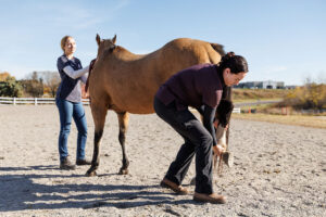
Equine Ringbone: Process, Progress and Prognosis
Here’s how veterinarians and farriers diagnose and manage this degenerative arthritic condition.

Here’s how veterinarians and farriers diagnose and manage this degenerative arthritic condition.

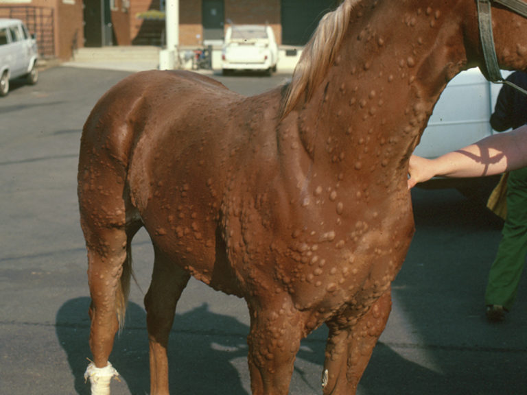
Find out what conditions, beyond lameness, a veterinarian might uncover during a prepurchase exam.

Find out what a veterinarian might look for when examining a horse that loses his balance after jumping a fence.

Two veterinarians share how they diagnose, treat, and rehab back-sore horses.

Radiographs can advise buyers and their veterinarians about pathologies that might pose a threat to the horse’s future soundness.
Four diagnostic imaging experts share insight that can help owners and veterinarians with the diagnostic process.
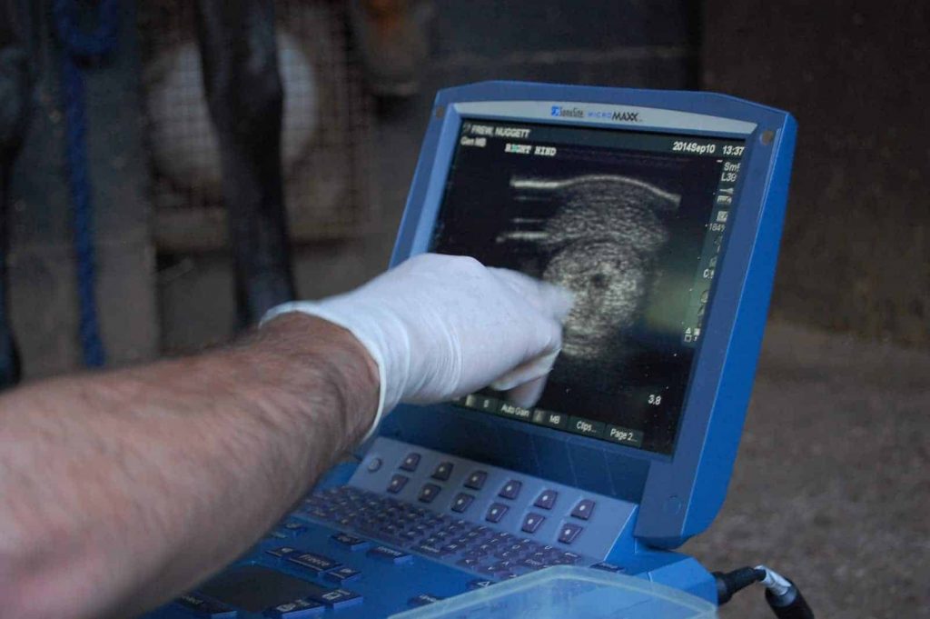
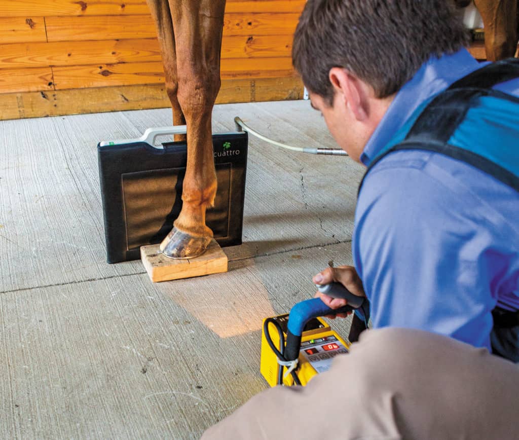
Diagnosing lameness in horses can be challenging, but veterinarians have an arsenal of imaging modalities available to help them make an accurate diagnosis.

Neck pain and pathologies are common in sport horses. It is important to combine results from clinical findings with imaging when making a diagnosis.
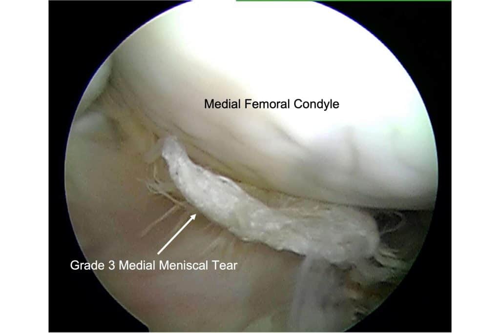
Equine meniscal injuries can cause severe pain and lameness, but if diagnosed and treated properly many horses can return to work.
X rays are valuable diagnostic tools for equine dental health that can reveal undiagnosed issues.

Veterinarians discuss how they radiograph, ultrasound, and treat neck problems.

Find out how to get athletic horses with injuries to the large, complex stifle joint on the road to recovery.
A veterinary podiatrist explains how X rays can be a useful tool for managing horses with chronic laminitis.

What can an owner do to control a horse’s foot pain? We turned to two equine veterinarians that spend a lot of time managing horses’ feet to find out.

Though it goes by several names, the condition is common among horses and frustrating for owners, farriers, and veterinarians alike.

MRI has helped veterinarians recognize new pathologies within horses’ hooves as well as learn more about existing lesions.