Can Hoof Wall Radiographs Help Identify Early Laminitis?
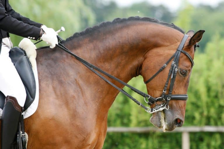
Radiographing deep hoof wall layers, where laminitic changes occur first, could be helpful in early clinical assessment.

Radiographing deep hoof wall layers, where laminitic changes occur first, could be helpful in early clinical assessment.

Improved diagnostics and more promising treatments are putting many foot-sore horses back to work.
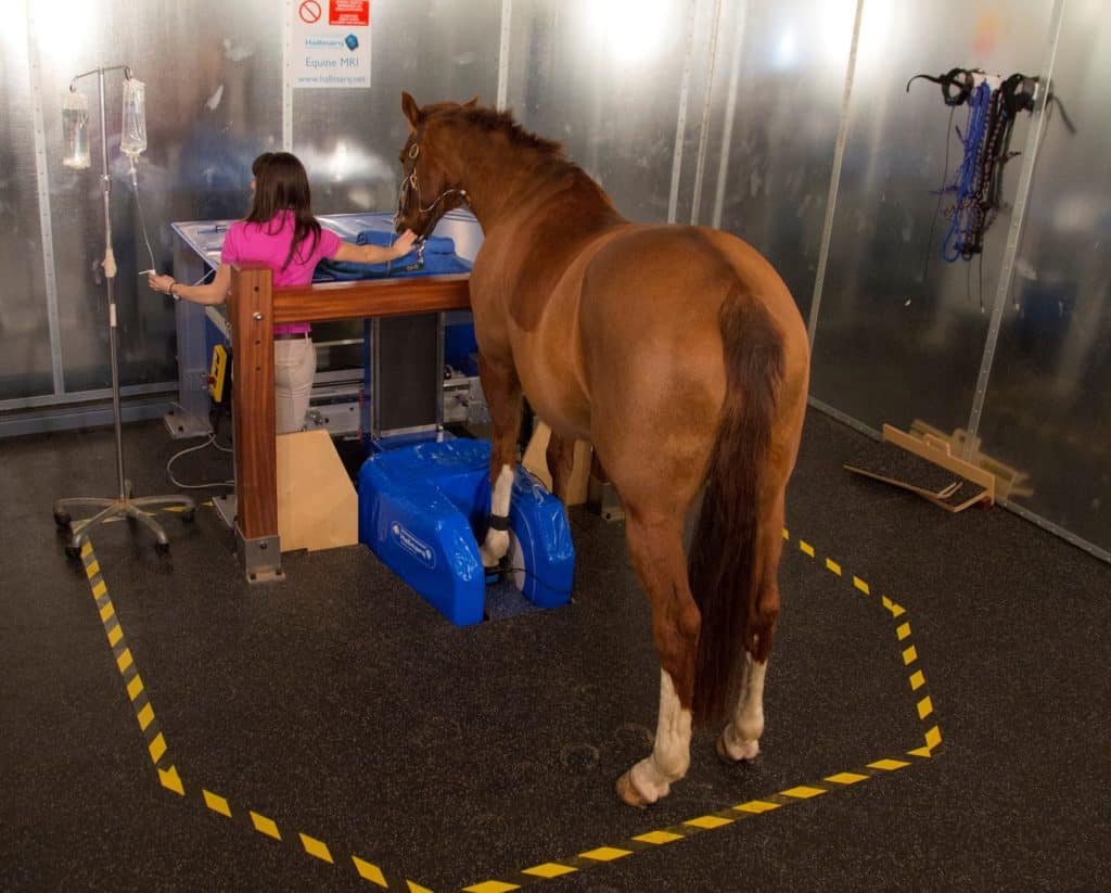
The four-robot system can perform multiple modalities and will be used in conjunction with a high-speed treadmill.

Radiographic abnormalities don’t always result in diminished performance; however, many do. Here’s what to know.
The horse’s lower limb is subject to a multitude of injuries that can baffle even the most veteran veterinarians.

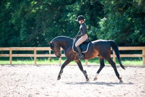
Poor performers can be a diagnostic challenge. Here’s what veterinarians will look for when examining these horses.
Are CT, MRI, and X ray clear as mud? Learn about the appropriate uses for these imaging modalities and more.
Of the 375 respondents, 201 (54%) said the most recent imaging modality used on their horses was radiography.

For the most part, the adverse reactions were mild and self-limiting. But some serious ones did occur.

Ultrasound is a useful method for diagnosing carpal sheath effusion, which is often associated with soft tissue damage.
Topics will include lower-limb lameness, ophthalmology, ambulatory practice, and poor performance.

Using proper diagnostic technique and good quality radiographs is critical when diagnosing these fractures in foals.

Review the options vets have for looking inside horses’ bodies to see what’s causing a limp, swelling, or pain.

A veterinarian weighs in on radiographs, nuclear scintigraphy, and standing MRI for diagnosing subtle lamenesses.
Radiographs (X rays) and low-field MRI appear to be useful tools for diagnosing early-stage arthritis.
The field protocol is designed to be low-risk, efficient, economical, and effective in identifying lesions.