Diagnosing Colic in a ‘FLASH’
Learn how vets use a technique called FLASH, a targeted abdominal ultrasound examination, to diagnose colic.

Learn how vets use a technique called FLASH, a targeted abdominal ultrasound examination, to diagnose colic.
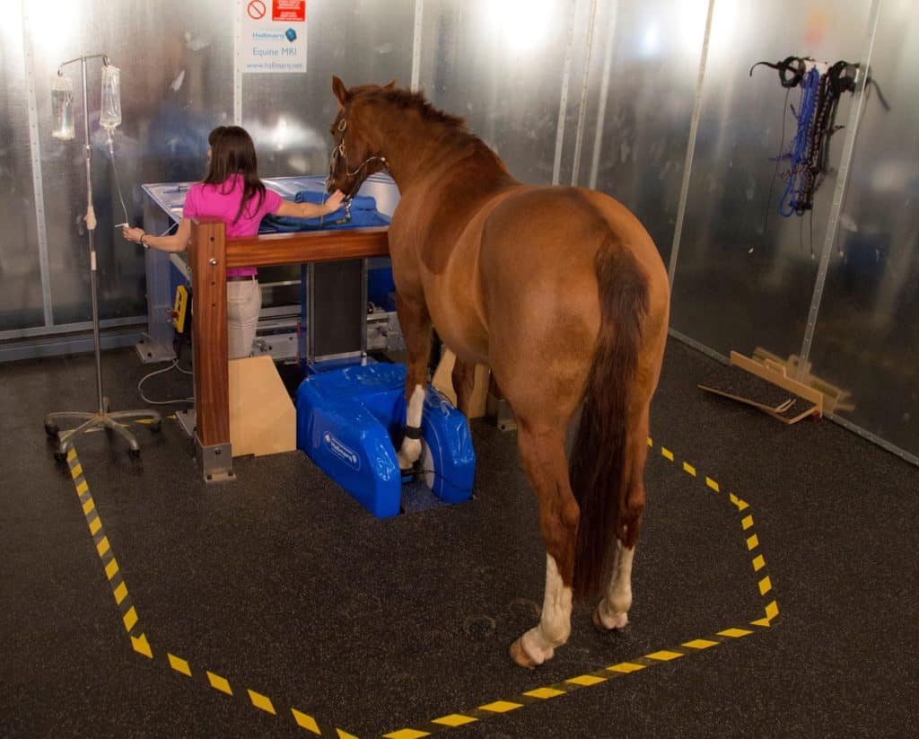
Are CT, MRI, and X ray clear as mud? Learn about the appropriate uses for these imaging modalities and more.

Of the 375 respondents, 201 (54%) said the most recent imaging modality used on their horses was radiography.

Take a behind-the-scenes look at the goings-on of a U.K. vet clinic and its differences from ones stateside.

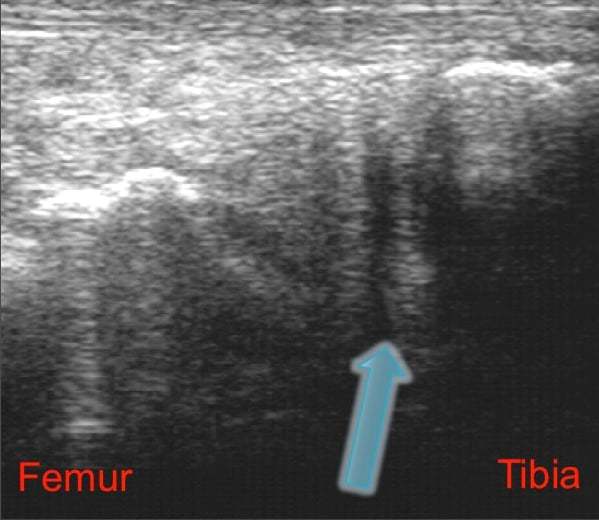
Ultrasound is a useful method for diagnosing carpal sheath effusion, which is often associated with soft tissue damage.
Researchers determined that using both modalities can help vets increase their diagnostic and prognostic capabilities.

A veterinarian weighs in on radiographs, nuclear scintigraphy, and standing MRI for diagnosing subtle lamenesses.

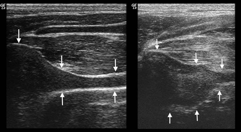
Vets can image the cecal mesentery to garner information that could lead to a lymphadenopathy diagnosis.
Reef will explain why ultrasound is critical to assessing foal viability during a high-risk pregnancy.

Colors displayed by Doppler ultrasonography could help vets better follow tendonitis’ healing processes.
The position will fund Cheetham?s research into diagnosing and treating recurrent laryngeal paralysis.

Researchers tested Doppler ultrasonography to detect vasoconstriction due to endophytic alkaloid consumption.
Researchers identified what ultrasound settings achieve adequate temperature increases to be therapeutic.

One veterinarian described the steps involved in conducting an ultrasound examination of the equine lungs.
Ultrasonography could be a valuable adjunct tool for diagnosing equine arytenoid chondritis.
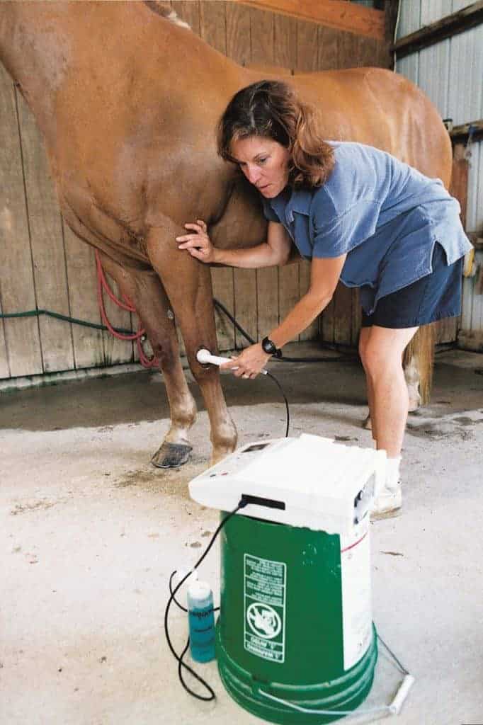
Ultrasound can be a valuable aid to practitioners when assessing the cause and severity of colic.
Notifications