Fetlock Injuries in Racehorses
Learn about the signs, diagnosis, and management of repetitive stress-related fetlock injuries in racehorses.

Learn about the signs, diagnosis, and management of repetitive stress-related fetlock injuries in racehorses.

Of the 359 respondents, 101 (28%) said their hoof care professionals use radiographs to make trimming/shoeing decisions.

Researchers focused on the effect of collected and lengthened paces in young and mature dressage horses.

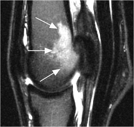
Vets can use MRI to help diagnose injuries, select treatments, monitor healing progress, and determine prognosis.

The digital cushion, located in the rear of the hoof, protects the foot’s complex bone and soft-tissue structures.

PET scans revealed lesions in bony and soft tissue, some of which weren’t visible on other imaging modalities.

Tailored rehab plans and frequent veterinary checks are just two pieces of the rehabilitation puzzle.
Read the top tweets and take-homes from Rood & Riddle Equine Hospital’s client education seminar.
Vets use MRI to identify issues and prescribe targeted treatment to give the horse the best chance at returning to work.

A hereditary disease–skeletal atavism–leads to disturbed skeletal development and usually requires euthanasia.
Researchers found that MRI images of bone thickness could provide critical information about fracture risk.

Veterinarians use imaging technologies to evaluate, diagnose, and ultimately treat lameness in the lower leg and foot.
Take a look at the evolution of diagnosing distal limb lameness in sport horse practice with Dr. Brendan Furlong.
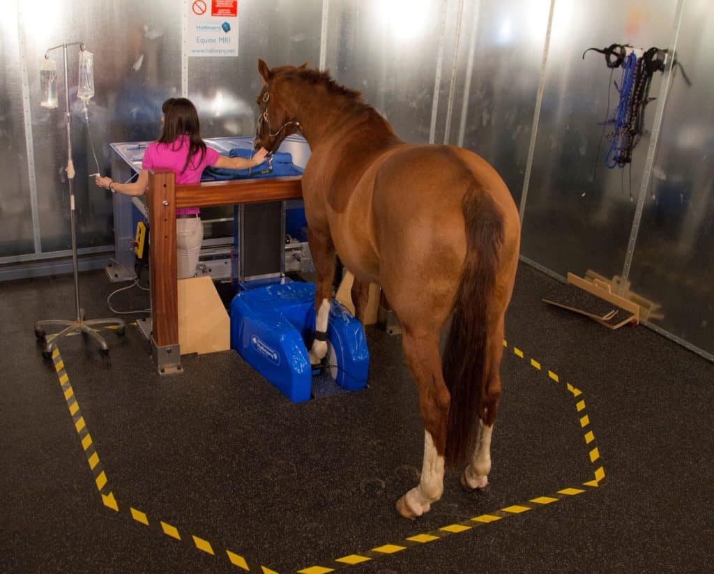
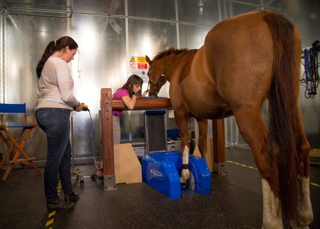
Researchers found that standing MRI is effective for viewing the difficult-to-image ligaments in the horse’s lower legs.

Horses’ knees are prone to both congenital and acquired lameness problems. Here’s what you need to know.

The team tested how much pressure compression and standing wraps exert–and with what consistency–on horses’ legs.