Ultrasound and sMRI Imaging of the Equine Foot Compared
Belgian researchers use both modalities to investigate the causes of foot pain in 30 horses referred to the equine hospital for forelimb lameness.

Belgian researchers use both modalities to investigate the causes of foot pain in 30 horses referred to the equine hospital for forelimb lameness.

Though it goes by several names, the condition is common among horses and frustrating for owners, farriers, and veterinarians alike.

MRI has helped veterinarians recognize new pathologies within horses’ hooves as well as learn more about existing lesions.

Diagnostic imaging technology has improved tremendously in the past few decades, with several effective options to choose from. Learn about the machines and technologies your veterinarian can use to look inside your horse, including MRI, CT, PET scans, and more.

Read about three real-life examples of equine athletes that made full recoveries from their injuries, including their diagnostic challenges, rehab modalities, and recovery details.

Researchers described normal pituitary gland appearance on MRI. Their findings might help veterinarians identify PPID in horses and start treatment earlier.

Computed tomography (CT) and magnetic resonance imaging (MRI) are two diagnostic imaging methods veterinarians can use to capture images of structures within your horse’s body. Learn more in this visual guide!

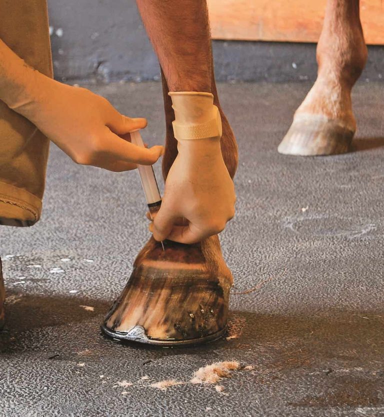
Lame horse? Advanced diagnostic and management strategies for navicular syndrome have improved long-term outcomes.

To help horse owners better understand MRI, we’ve scoured our archives and collected 10 resources about this diagnostic modality and some of the ailments it can help identify, all available to you free on TheHorse.com.
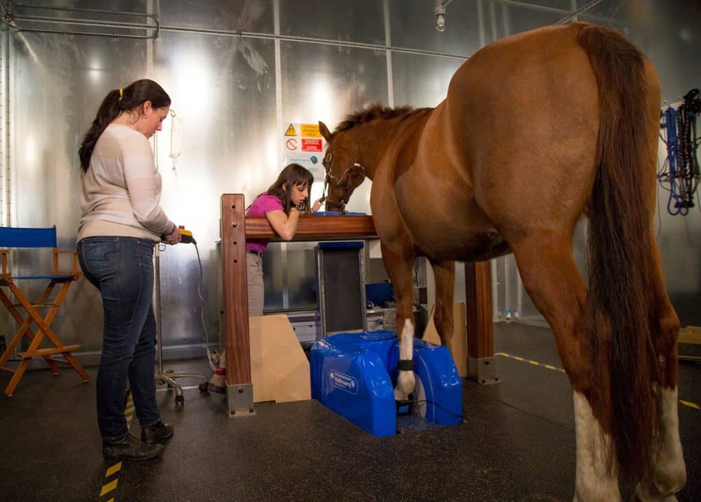
MRI has become easier to use and more accessible. Using this diagnostic tool early leads to improved outcomes for lame horses. Sponsored by Hallmarq Veterinary Imaging.
MRI exams are more expensive than most other diagnostics. But with better associated recovery rates, it might be a worthwhile investment, say researchers.

One veterinarian weighs the pros and cons of available imaging modalities when it comes to diagnosing common fetlock injuries.

One practitioner describes her diagnostic imaging decision-making process when assessing Western performance horse soundness during prepurchase exams.
Here’s what we know and are learning about defining, diagnosing, and treating this painful foot disease.

Researchers found that high-field MRI provided highly detailed and anatomically accurate images of equine stifle soft tissues.

MRI revolutionized the way veterinarians diagnose problems with the equine podotrochlear apparatus. Dr. Kyla Ortved explains its importance and when it’s worth the expense.