A Guide to Equine Diagnostic Imaging

Learn about machines and technology your veterinarian can use to look inside your horse
Over the past few decades manufacturers have introduced new equine diagnostic techniques and enhanced existing imaging modalities. Today’s equine veterinarians have many tools at hand to help pinpoint problem areas and diagnose issues. Some of these modalities are portable enough to take on a farm call, while others are only accessible at clinics or hospitals.
You have the old standbys, such as radiographs (X rays) and ultrasound, plus newer technologies, including MRI, CT, and PET scan, among others. In this article we’ll review the machines your veterinarian might use to look inside your horse and why.
Radiographs
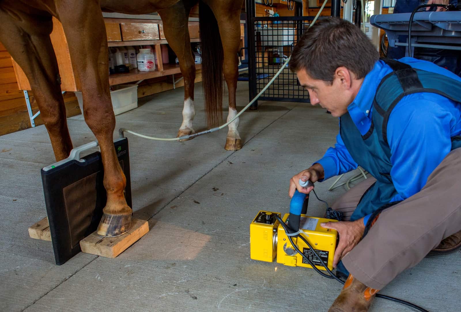
Radiograph technology is the oldest imaging modality, having been around for more than a century. It has undergone a dramatic evolution in the past 20 years, from film vets had to develop back at the clinic to nearly instantaneous high-quality images captured with wireless equipment practitioners can use anywhere.
“The earliest digital X ray systems were of considerably lesser quality than what is available now, and this is true of all digital and computer technologies,” says Sarah Puchalski, DVM, Dipl. ACVR, owner of Puchalski Equine Diagnostic Imaging, in Petaluma, California. “They keep evolving and are leaps and bounds ahead of where they once were. A lot of the new systems are battery-operated and completely portable.”
Today most veterinarians use these digital radiograph machines for an immediate look at the horse’s internal structures. As an added bonus, says Mark Reilly, DVM, Dipl. ABVP, founder of South Shore Equine Clinic and Diagnostic Center, in Plympton, Massachusetts, veterinarians can enhance (level, enlarge, invert) the images and share them almost instantly through email.
“At the farm we can get good-quality X rays for the lame horse or do prepurchase exams,” says Puchalski. “For emergency situations on the farm, stallside digital X rays can look at fractures, wounds, etc. on a limb but are not as useful for things like colic or pneumonia where we need abdominal or thoracic (chest) views.”
Veterinarians need high-energy X rays, which portable machines cannot deliver, to view parts of the neck, back, and abdomen and thicker, deeper areas of the body, she says. The horse typically needs to go to a clinic or hospital for these.
Ultrasound
Veterinarians commonly use this portable modality to conduct reproductive evaluations, assess pregnancy, and evaluate injured tendons and ligaments. Ultrasound can reveal details about soft tissues that X rays cannot.
“Ultrasound is complementary to X rays, and generally both are used to obtain a diagnosis,” says Puchalski. For example, if a horse is lame, when the veterinarian figures out which part of the leg is causing the problem, he or she might use radiographs and ultrasound to fully evaluate the injury.
“Ultrasound is useful for looking at soft tissues and bone surfaces,” she says. “It can be used to evaluate the pelvis, for instance, whereas X rays of the pelvis are difficult because (the soft tissue covering) this area is too thick. If the horse had a fall or trailer accident and pelvic fracture is suspected, we have difficulty getting radiographs, but we can use ultrasound to evaluate the bone surface and see if a fracture is visible.”
Veterinarians can also use the ultrasound probe to guide needle placement, which helps make administering therapeutic agents more accurate.
In the past 20 years ultrasound has undergone dramatic digitization, evolution, and improvement. “Now we have extremely high-detail probes with excellent resolution so we can see different things—and very small things—very clearly,” says Puchalski.
As a result, veterinarians are learning to use ultrasound to look at eye problems, wounds, masses, and more, says Reilly.

Magnetic Resonance Imaging
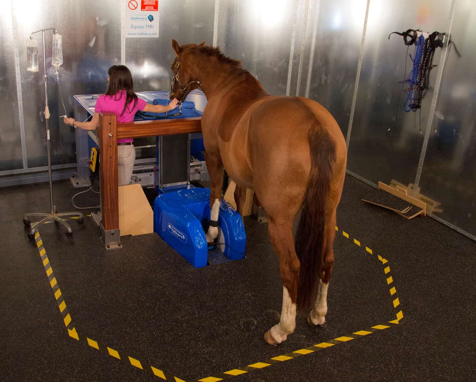
With MRI, a magnetic field and computer-generated radiofrequency waves create detailed images. “This takes very specialized equipment,” says Puchalski. “For humans, MRI is considered a gold standard for the diagnosis of musculoskeletal problems because it will image bone, soft tissue, and fluid (such as synovial fluid in a joint). These images provide high detail and cross-sectional slices of the tissue. We can see slices through the bone, tendons, and ligaments, rather than just looking at the whole bone.”
“(MRI) completely changed our understanding of navicular disease,” she says, referring to cases of heel pain related to the navicular bone and its associated structures.
Adds Reilly, “MRI has revolutionized diagnostics and treatment of foot lameness because we can see inside the hoof,” with detail, something that is difficult to impossible with other imaging modalities.
Scans can be done standing, with the horse sedated, or lying down, with the horse anesthetized. “The disadvantage to general anesthesia is risk for the horse, which has nothing to do with the MRI, but this is unappealing to many horse owners,” especially for checking a mild lameness, she says.

An MRI’s magnet strength can be either low-field or high-field. “All standing MRI units are low-field, so those images have less detail than high-field MRI and don’t image cartilage as well,” says Puchalski. Veterinarians can only perform standing MRI on horses’ lower limbs, which is where the vast majority of lamenesses occur, she says.
“For a brain problem or something else regarding the head, the only option would be to perform the study with the horse lying down and under anesthesia,” she says.
Horse owners should weigh the pros and cons of each type of MRI based on their individual situations. “It’s helpful to have a conversation with your veterinarian to determine what’s best,” says Puchalski. “Your choice may also depend on where you live, because some regions have many options and others are limited.”
Computed Tomography (CT)
Computed tomography is an imaging procedure that uses special X ray equipment to create detailed pictures of areas inside the body. It was previously termed computerized axial tomography (CAT) scan. “This is essentially cross-sectional radiographs and very useful, providing excellent, high-detailed images for bone and fair to good images for soft tissues. In general, it is not as good as MRI for viewing soft tissues,” says Puchalski.
Depending on the clinic, veterinarians can perform CT scans with the horse standing and sedated or under general anesthesia.

“Historically, one of the challenges with CT scanners is they have a round opening that can almost always fit the limbs but might not physically accommodate larger parts of the horse’s body, such as the neck or upper limbs,” says Puchalski. “Within the past five years, the medical imaging companies have made large machines designed for bariatric medicine, and there are now a number of ‘large-bore scanners’ in equine practice. We can get the horse’s entire head and neck into these machines.”
Veterinarians often use CT to diagnose dental or sinus disease, using it “to look at unusual head swellings or sinus swellings or fractures,” says Reilly. “With X rays sometimes it’s hard to tell, with all the fluid and disturbance that occurred in the skull, whereas the CT defines it nicely.”
Depending on where you live, you may have access to the large-bore scanner technology. Puchalski says there are two available in California, for instance.
Some equine clinics also have robotic cone-beam CT scanners that move around the standing horse. “Several places have one of these, including New Bolton Center, but there are not many yet in private practices,” says Reilly. “It is relatively inexpensive for the client because it’s quick; the horse just stands there sedated and the machine goes over both sides of the horse and moves around to get a lot of images.”
“The technology is still young, however, and image quality is not as good as conventional scanners,” adds Puchalski.
What’s This Going to Cost?
Some imaging modalities are more expensive than others, and prices vary widely between practices and regions. “Digital X rays probably run between $50 and $60 per view for a particular site,” Mark Reilly, DVM, Dipl. ABVP, founder of South Shore Equine Clinic and Diagnostic Center, in Plympton, Massachusetts, says of his practice. “We rarely take less than two views and often take six to eight views. If you are doing prepurchase exams or advanced imaging with multiple sites, sometimes these are packaged.”
He says an ultrasound typically costs a few hundred dollars. “In our area, it runs around $300, and this is generally based on the technology and the price to purchase it,” he says.
Bone scans can run anywhere from $1,500 to $2,500 once you factor in hospitalization, sedation, catheterization, and more, he says.
“CT usually requires general anesthesia, if the horse is lying down, and will run about $2,000,” Reilly says. “If they can do it standing it will probably be less than $1,000—maybe about $700 or $800 and include the hospitalization. A standing MRI will be about $2,200, which includes the interpretation. I’ve seen anesthetized MRI costing $2,500 to $2,700 or more.”
—Heather Smith Thomas
Nuclear Scintigraphy (Bone Scan)
Veterinarians can perform bone scans in standing sedated horses at the clinic. “A radioactive substance is injected through a catheter placed in the jugular vein,” says Reilly. “This radioisotope has a very short half-life of six hours (i.e., half of the radioactivity will be gone in six hours). Within a few hours of the injection, the horse is moved to a room where a large gamma camera is moved around the horse, and it records where the body has increased amounts of uptake of the substance. These areas are commonly known as hot spots—areas of increased bone activity (or soft tissue inflammation or cell turnover). Abnormal areas can then be further imaged with another modality (e.g., X rays, ultrasound, MRI) to determine the extent of the injury.”
Thus, this technology is very useful for finding the general location of a problem.
The radioisotope uptake, says Puchalski, “might be because of a fracture or bone inflammation, osteoarthritis, or a number of other things that cause the bone to be remodeling. Wherever bone cells are turning over (destruction/resorbing of the mineral matrix and new material being laid down) the radioactive tracer will accumulate.
“These cameras are fairly large—up to 14 by 17 inches—and map as much of the skeleton that will fit on the detector,” she adds. “It shows the areas with high activity and is most useful for screening when a horse has a nonspecific musculoskeletal problem that we haven’t been able to pinpoint. There may not be a detectable specific lameness, but the horse has poor performance.”
Veterinarians can also use scintigraphy to look at large, difficult-to-radiograph areas such as the pelvis, neck, and back. “We also use this technology when a horse has multiple-limb lameness, such as a hind limb and a front limb,” says Puchalski. “We can look at the whole skeleton at the same time.”
Before sending a horse home from the clinic post-scan, the veterinarian should measure the horse’s radioactivity to make sure it has returned to a safe level. “The downside of using CT or nuclear scintigraphy is that it involves radiation,” says Reilly. “There are obviously some human health hazards with the radiation. With a bone scan, the horse must have a gamma count (performed on blood) before it goes back to the owner, to make sure it is not urinating radioactive material.”

Positron Emission Tomography (PET)
This new equine imaging technology in development at the University of California, Davis (UC Davis), uses a radioactive tracer to show how tissues and organs are functioning.
“The PET scan can be thought of as cross-sectional nuclear scintigraphy, where slices of tissue are evaluated. This is sometimes done in concert with CT scanning,” says Puchalski. “The radioactive material can show areas where high cell metabolism is taking place in bones, using a slightly different biological mechanism to create the image.”
While this technology is still very new in horses, and veterinarians don’t have much experience with it yet, she says it might be useful as a screening tool, such as for soundness and safety in racehorses. In fact, UC Davis has partnered with Santa Anita Park to install a PET scanner specifically designed for equine limbs, so veterinarians have another way to look for pre-existing injuries.
The Veterinarian’s Role
When discussing all the cool, high-tech machines that can view inside horses’ bodies, we can’t forget about the human element.
“The most important part of any diagnostic workup is good veterinary oversight—a person who knows the horse and what is expected of it, along with intimate knowledge of the clinical problem,” says Sarah Puchalski, DVM, Dipl. ACVR, owner of Puchalski Equine Diagnostic Imaging, in Petaluma, California. “All of these diagnostic imaging technologies are useful, but if you don’t have an appropriate workup and someone who can take that information and apply it back to the horse for proper treatment, it won’t do that much good.
“Sometimes people view diagnostic imaging like a vending machine, where you stick the horse in and then the answer comes out, but it’s not that simple,” she continues. “It’s usually just one component of a complicated workup.”
A common misconception among horse owners is that they can have their veterinarians perform an MRI, for example, and it will give them all the information they need. “That’s not the whole picture (it still needs to be interpreted and applied to the horse), and sometimes it’s only a small component of the cost involved when dealing with the horse’s problem,” says Puchalski. “In many circumstances, the cost of an MRI may be the least of the total cost to diagnose and treat the horse and, in fact, spending money on expensive diagnostics early in the process can actually save the horse owner money in the long run, by ensuring that the treatment is correct for the horse’s condition.”
—Heather Smith Thomas
Endoscopy
Veterinarians can pass a 1-meter fiber-optic endoscope through the nostril and into the airway to view the upper respiratory tract. It lets them visualize the throat (pharynx, larynx, guttural pouches, epiglottis, and soft palate) and see into the trachea. Longer scopes can reach as far as the horse’s stomach to look for ulcers.
The former “allows us to see abnormal function of the airway, as well as any exudate (pus), blood, or foreign bodies within the upper respiratory tract,” says Reilly. “The endoscope has a biopsy channel, which allows a variety of diagnostic tools (cytology brushes, probes, biopsy forceps) to be passed to get a sample of any exudate, remove a foreign body, or potentially perform airway surgery.”
He adds that most horses tolerate endoscopic procedures well, with simply a twitch or a lip shank and sometimes light sedation.
Venogram
A venogram is a radiograph taken after injecting contrast material (dye) into a vein to show how blood flows through it. “This is useful for (working up cases of) laminitis,” says Puchalski.
It enables the veterinarian to view the blood vessels inside the hoof capsule and, therefore, the status of the foot’s blood supply, which does not show on normal radiographs. If there is swelling within the hoof capsule or the coffin bone is displaced (rotated or sinking) due to laminitis, she explains, the blood vessels can become occluded or damaged, diminishing or curbing blood flow.
Venograms allow veterinarians to determine what is wrong and where, and come up with an effective treatment plan.
Take-Home Message
Diagnostic imaging technology has improved tremendously in the past decades, with several effective options to choose from. Portable on-the-farm diagnostics primarily include radiographs and ultrasound, while more highly specialized and expensive diagnostics, such as scintigraphy, MRI, or CT, must be done in a hospital setting.
“There are a few new, developing technologies that may one day become stallside but currently are not,” says Puchalski.
What you and your veterinarian select depends on your horse’s injury or health condition and the imaging technology available in your region. Costs also vary, depending on the practice or hospital and its geographic location.
Written by:
Heather Smith Thomas
Related Articles
Stay on top of the most recent Horse Health news with


