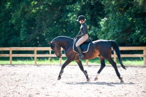
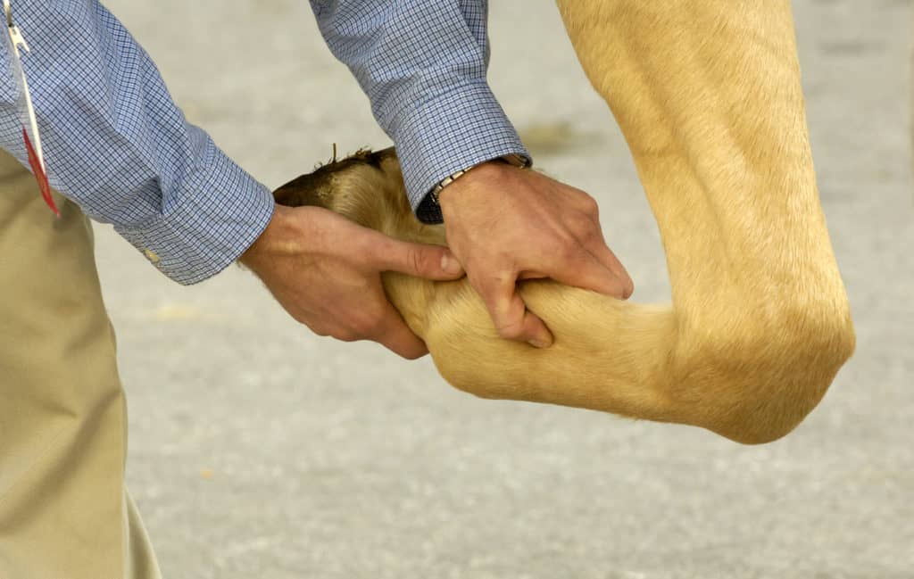
The Horse’s Skeleton: Forelimbs
Learn about the structure and function of your horse’s forelimbs with Dr. Roberta Dwyer of the University of Kentucky’s Gluck Equine Research Center.
Prevention, diagnosis, and treatment of leg lameness
Learn about the structure and function of your horse’s forelimbs with Dr. Roberta Dwyer of the University of Kentucky’s Gluck Equine Research Center.
It’s important an owner knows how to help his or her injured horse until the veterinarian arrives.
Dr. Vernon Dryden of Rood & Riddle Equine Hospital demonstrates navicular syndrome diagnosis in a 16-year-old Quarter Horse.
It has been suggested that 60% of equine lameness problems are related to osteoarthritis, which stresses the importance of advancements of both medical & surgical treatment options.
A joint is defined as an anatomic union or junction between two or more bones. There are three basic types of joints in the horse: Synovial, fibrous, and cartilaginous.
Equine joint supplements have been purported to decrease inflammation, increase mobility, and more. This guide to equine joint supplements includes a seven-step quality evaluation process and tips for choosing a safe and effective product.
Equine joint injections can help veterinarians diagnose lameness or medicate a horse’s painful joint.
Equine lameness, defined as a deviation from a normal gait, is an indicator of a structural or functional disorder of the musculoskeletal system (the limbs or spinal column) that is noted while the horse is either moving or stationary.
Pinpointing lameness in horses is crucial before proper treatment can be prescribed. There are several approaches to lameness examinations and diagnostic methods, which will be partly determined by the age of the horse and what it does for living.
In a retrospective study of 118 French Trotters, a team of veterinarians found that horses with back pain had more severe and localized lesions identifiable on X ray than horses with no evidence of back pain, but even pain-free horses had lesio
Foot lameness in horses was the topic of two separate Table Topics during the 2009 American Association of Equine Practitioners (AAEP) Convention in Las Vegas, Nev., in early December. About 100 veterinarians attended each session.
A
Learn about the normal functions of bones, cartilage, soft tissues, and fluids within the fetlock joint.
Strategies to identify specific problems in a horse with indistinct or unusual gait deficits, particularly if
A recent study of coffin bone (distal phalanx) fractures in foals found they were far from rare. In fact, all 20 of the Warmblood foals in the study (all foals on a particular farm in one season) had fractures at some point in their first year of life.
New imaging technologies, such as MRI, CT, digital ultrasound, and nuclear scintigraphy, have helped us understand anatomy better and improved our ability to visualize joint injury. As a result, we’re now able to utilize nonsurgical joint therapies.
Knowing how the hoof is built and what it is capable of can help you understand what it needs to stay healthy and recover if compromised.
Stay on top of the most recent Horse Health news with
"*" indicates required fields