Poll Recap: Getting a Closer Look
Of the 375 respondents, 201 (54%) said the most recent imaging modality used on their horses was radiography.

Of the 375 respondents, 201 (54%) said the most recent imaging modality used on their horses was radiography.
The practice, located in Petaluma, California, also offers equine sports medicine and rehab services.
Cornell Ruffian Equine Specialists hopes to use the state-of-the-art imaging system to help assess lameness.
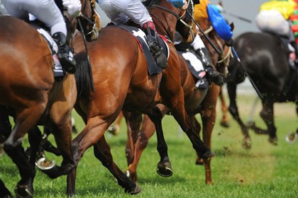
Researchers say MRI might be useful as a screening test to identify racehorses at increased risk of fetlock fracture.
Topics will include lower-limb lameness, ophthalmology, ambulatory practice, and poor performance.

Review the options vets have for looking inside horses’ bodies to see what’s causing a limp, swelling, or pain.

Horses with concurrent deep digital flexor tendon lesions were four times more likely to become lame again post-surgery.

Cornell Ruffian Equine Specialists signed an agreement to install the unit at its facility near Belmont Park.

A veterinarian weighs in on radiographs, nuclear scintigraphy, and standing MRI for diagnosing subtle lamenesses.
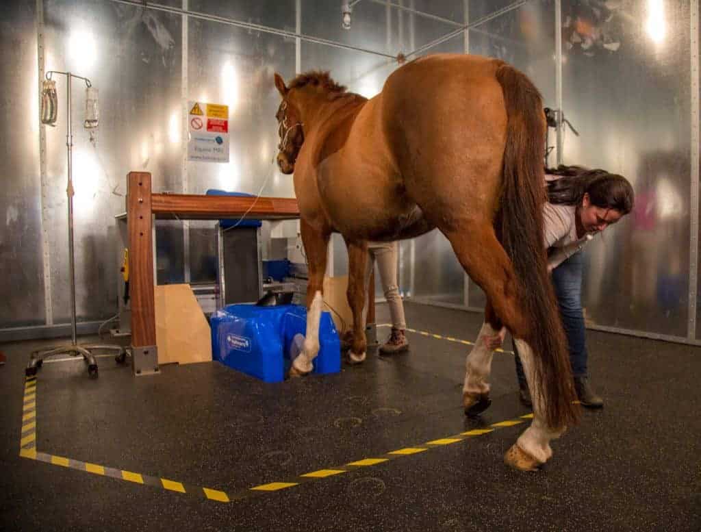
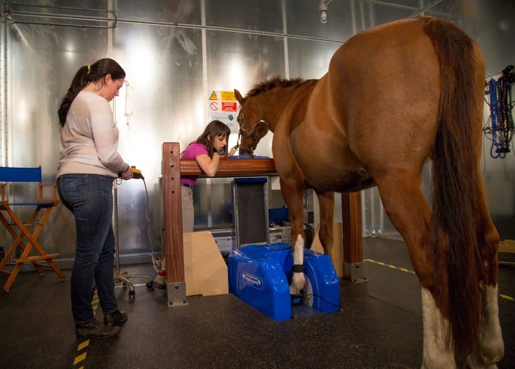
Dr. Rachel Buchholz explains the steps involved in having a standing MRI done on a horse.

The standing MRI is the first installed at a Thoroughbred racetrack or training center in North America, officials say.
Radiographs (X rays) and low-field MRI appear to be useful tools for diagnosing early-stage arthritis.
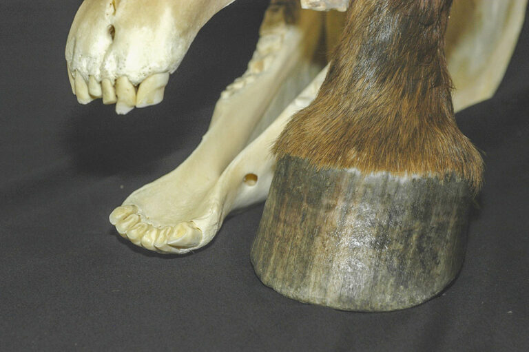
Learn about the standing MRI process step-by-step and take a look inside the equine foot.
Learn why magnetic resonance imaging (MRI) considered the “gold standard” for diagnosing equine lamenesses and get your questions answered about how MRI can help your horses during this live event.

Find out why standing MRI is useful for identifying complex issues in horse hooves and limbs.

Veterinarians have tools to help them make educated judgments about lamenesses, their causes, and prognoses.