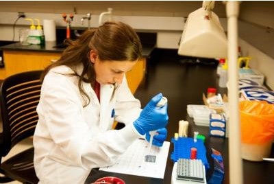
New Technology Speeds Animal Disease Diagnosis
Scientists are hailing a new technology called MALDI-TOF as a game changer for Kentucky animal health.

Scientists are hailing a new technology called MALDI-TOF as a game changer for Kentucky animal health.
How long will a horse with osteoarthritis remain sound after successful IRAP treatment?

Lavage systems placed in the upper and lower eyelids developed complications 12.8% and 22.2% of the time, respectively.

Learn how vets use a technique called FLASH, a targeted abdominal ultrasound examination, to diagnose colic.
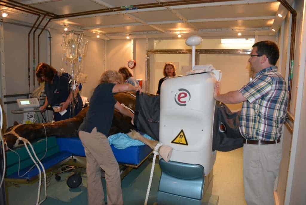
The new MRI replaces a previous system at New Bolton Center, installed in 2005 and used until recently.
Cornell Ruffian Equine Specialists, located next to Belmont Park, celebrated its first year in business on June 25.
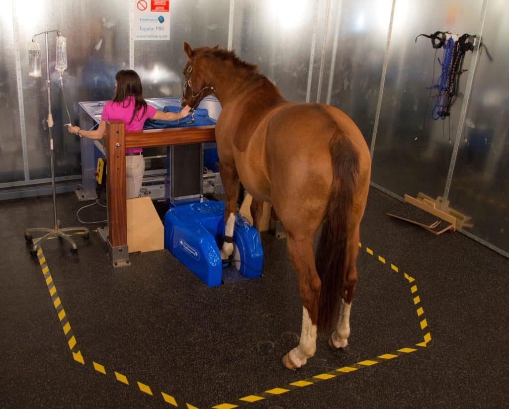
Are CT, MRI, and X ray clear as mud? Learn about the appropriate uses for these imaging modalities and more.
The MRI unit adds to the school’s imaging modalities, including a CT, radiography, nuclear scintigraphy, and more.

Of the 375 respondents, 201 (54%) said the most recent imaging modality used on their horses was radiography.
The practice, located in Petaluma, California, also offers equine sports medicine and rehab services.

Take a behind-the-scenes look at the goings-on of a U.K. vet clinic and its differences from ones stateside.
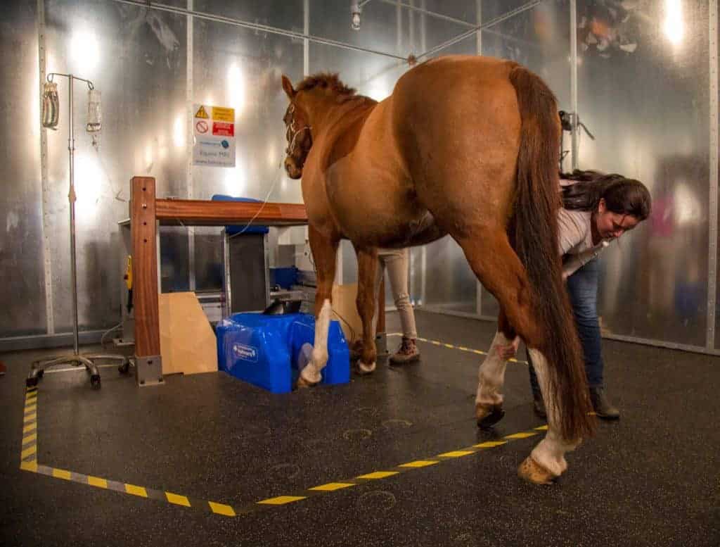
Cornell Ruffian Equine Specialists hopes to use the state-of-the-art imaging system to help assess lameness.

For the most part, the adverse reactions were mild and self-limiting. But some serious ones did occur.
The Veterinary Diagnostic Lab is in-the-know on animal diseases, including equine ones, around the state and country.
Equine Guelph and Dr. Judith Koenig received a donation help to purchase a lameness evaluation device.

Ultrasound is a useful method for diagnosing carpal sheath effusion, which is often associated with soft tissue damage.